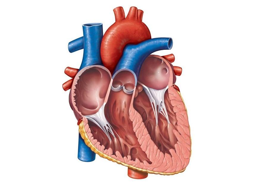
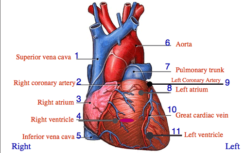
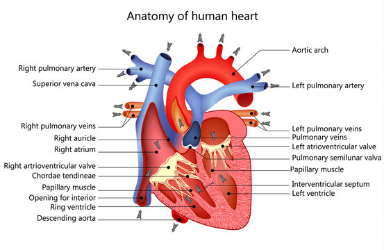
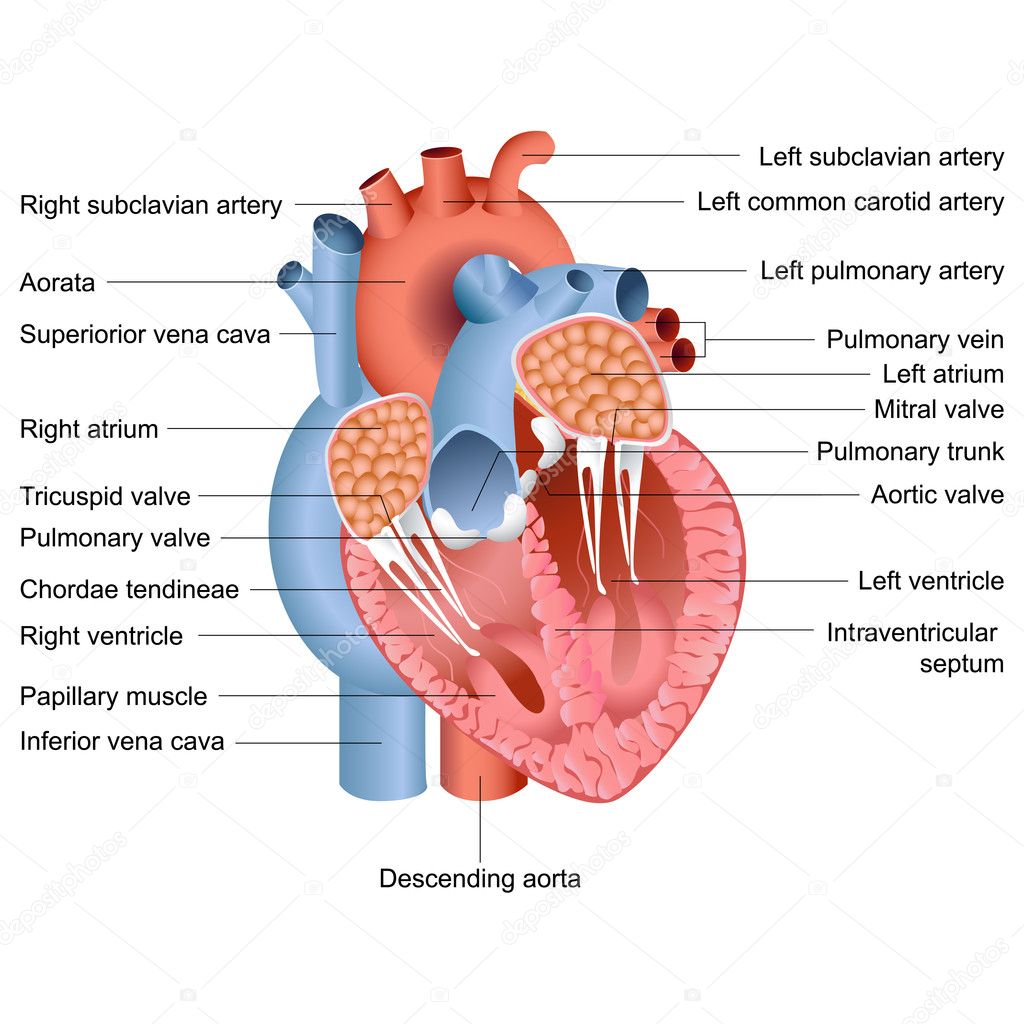
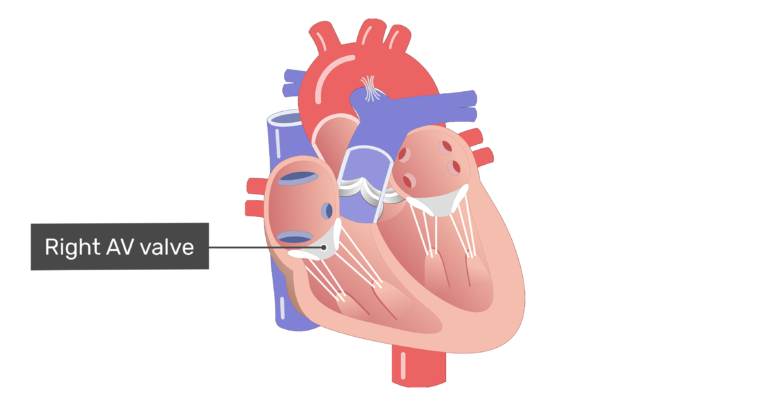
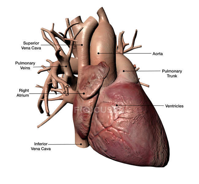
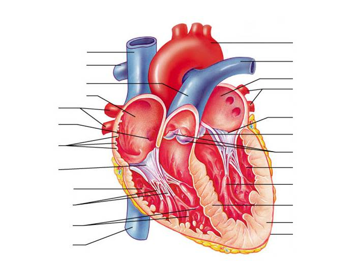
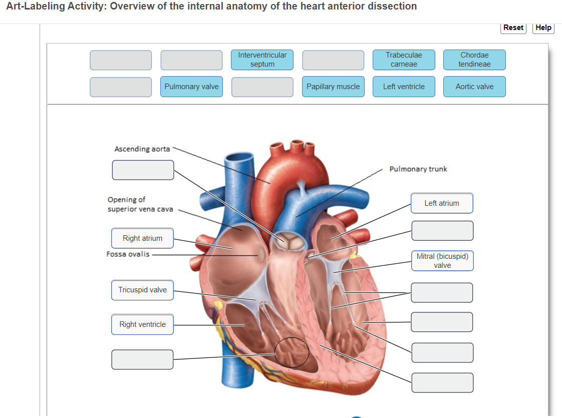
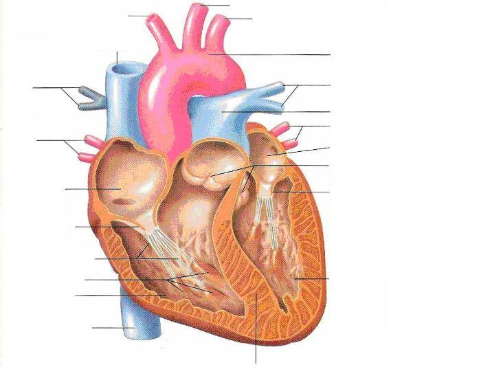
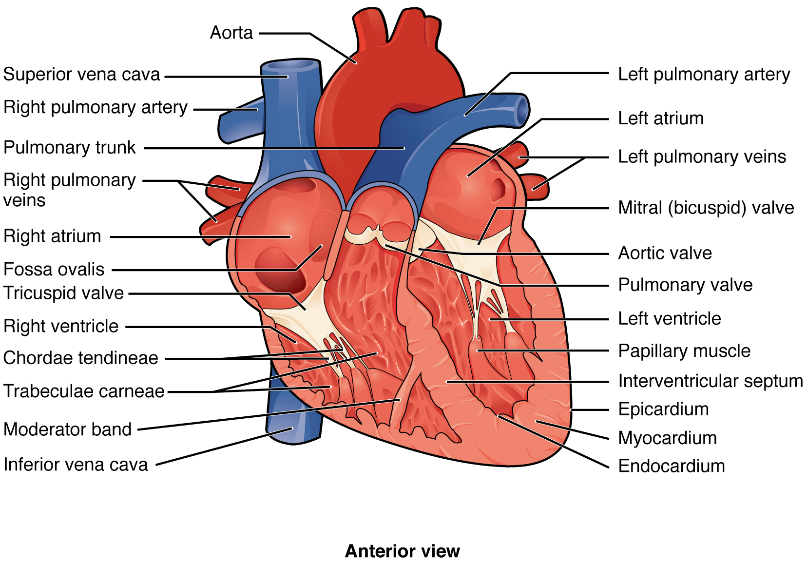
45 label the internal anatomy of the heart.
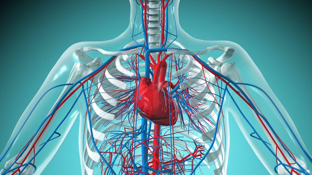
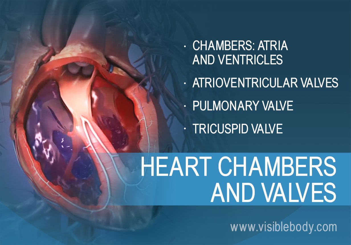
Internal structure of heart | Internal anatomy of the heart | Thorax ... Internally, the heart is divided into four chambers: . Two upper chambers known as atria: right atrium (RA) and left atrium (LA);; Two lower chambers called ventricles: right vetricle (RV) and left ventricle (LV).; Functionally, the heart consists of two pumps (right and left), each consisting of an atrium and a ventricle separated by an atrioventricular valve. Heart Information Center: Heart Anatomy | Texas Heart Institute A wall of muscle called the septum separates the left and right atria and the left and right ventricles. The left ventricle is the largest and strongest chamber ...
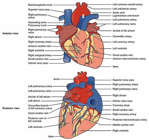
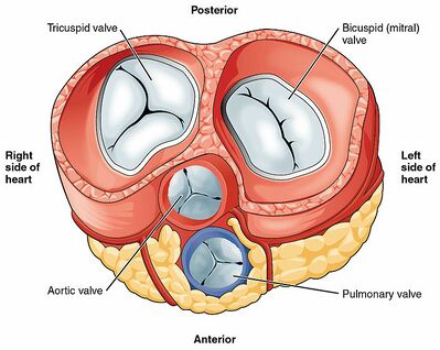
OpenStax AnatPhys fig.19.9 - Internal Anatomy of the Heart WebInternal Structures of the Heart. This anterior view of the heart shows the four chambers, the major vessels and their early branches, as well as the valves. The presence of the …
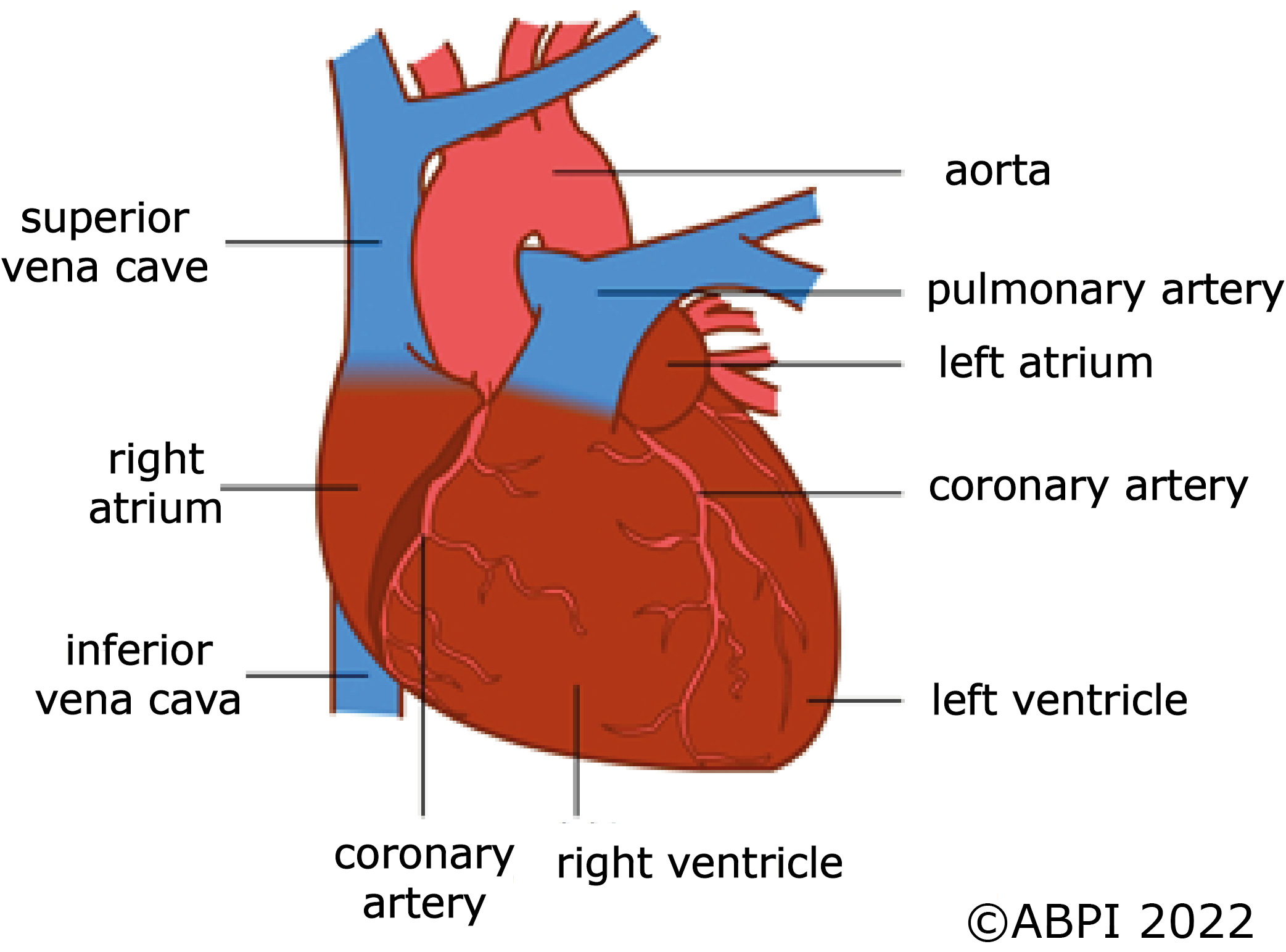
Label the internal anatomy of the heart.
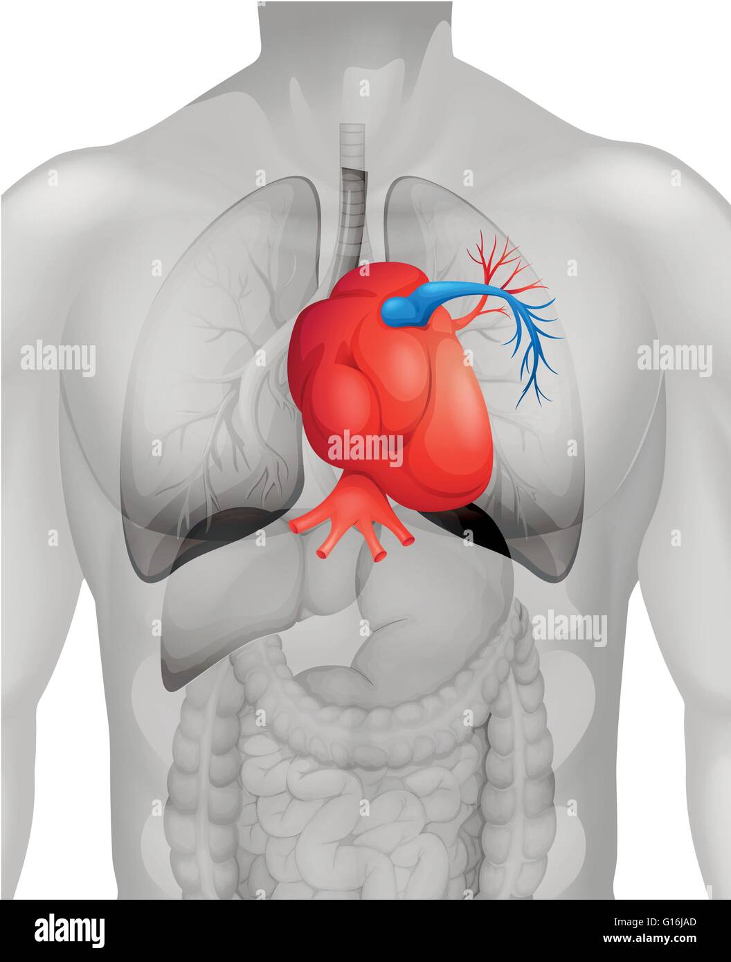
19.1 Heart Anatomy - Anatomy and Physiology 2e | OpenStax Location of the Heart. The human heart is located within the thoracic cavity, medially between the lungs in the space known as the mediastinum. Figure 19.2 shows the position of the heart within the thoracic cavity. Within the mediastinum, the heart is separated from the other mediastinal structures by a tough membrane known as the pericardium ... OpenStax AnatPhys fig.19.9 - Internal Anatomy of the Heart Internal Structures of the Heart. This anterior view of the heart shows the four chambers, the major vessels and their early branches, as well as the valves ... Layers of the heart: Epicardium, myocardium, endocardium - Kenhub The inner layer lines the heart chambers and is made of endothelial cells. Superiorly, is the second layer: a subendocardial connective tissue which is continuous with the connective tissue of the myocardium. Branches of the heart's conduction system are immersed into the subendocardial layer. Solidify your knowledge with our quiz:
Label the internal anatomy of the heart.. Heart Anatomy Labeling Game - PurposeGames.com Feb 22, 2022 · Heart Anatomy Labeling Game by htran9 4,007 plays 19 questions ~50 sec English 19p More 2 too few (you: not rated) Tries 19 [?] Last Played February 22, 2022 - 12:00 am There is a printable worksheet available for download here so you can take the quiz with pen and paper. Remaining 0 Correct 0 Wrong 0 Press play! 0% 08:00.0 Show More Heart Anatomy | Anatomy and Physiology II - Lumen Learning The majority of the internal heart structures discussed in this and subsequent sections are illustrated in Figure 8. While the bulk of the internal surface of the right atrium is smooth, the depression of the fossa ovalis is medial, and the anterior surface demonstrates prominent ridges of muscle called the pectinate muscles. The right auricle ... Chapter 20-Cardiovascular System Flashcards | Quizlet WebCorrectly label the following internal anatomy of the heart. b Place the labels in order denoting the flow of oxygenated blood through the heart beginning with the vessels that … Heart | Structure, Function, Diagram, Anatomy, & Facts heart, organ that serves as a pump to circulate the blood. It may be a straight tube, as in spiders and annelid worms, or a somewhat more elaborate structure with one or more receiving chambers (atria) and a main pumping chamber (ventricle), as in mollusks. In fishes the heart is a folded tube, with three or four enlarged areas that correspond to the chambers in the mammalian heart. In animals ...
Heart: Anatomy and Function - Cleveland Clinic Your heart walls are the muscles that contract (squeeze) and relax to send blood throughout your body. A layer of muscular tissue called the septum divides your heart walls into the left and right sides. Your heart walls have three layers: Endocardium: Inner layer. Myocardium: Muscular middle layer. Epicardium: Protective outer layer. The Anatomy of the Heart, Its Structures, and Functions - ThoughtCo The heart is made up of four chambers: Atria: Upper two chambers of the heart. Ventricles: Lower two chambers of the heart. Heart Wall The heart wall consists of three layers: Epicardium: The outer layer of the wall of the heart. Myocardium: The muscular middle layer of the wall of the heart. Endocardium: The inner layer of the heart. Label the heart — Science Learning Hub Web16. Juni 2017 · Label the heart. Interactive. Add to collection. In this interactive, you can label parts of the human heart. Drag and drop the text labels onto the boxes next to the diagram. Selecting or hovering over a … 6-5 The Internal Anatomy Of The Heart Answer Key PDF Internal Anatomy Of Heart - Mrs. Hille's FunZone. 6-5. THE INTERNAL ANATOMY OF THE HEART, continued 5. A large valve with three cup-shaped flaps lies between the right atrium and the right ventricle. (Note: To make the diagram easier to understand, the flaps have been drawn to look more like cups than they really would.)
The structure of the heart - Structure and function of the heart ... The heart is a large muscular pump and is divided into two halves - the right-hand side and the left-hand side. The right-hand side of the heart is responsible for pumping deoxygenated blood to... CH. 20 Assessment Flashcards | Quizlet Study with Quizlet and memorize flashcards containing terms like Correctly label the internal anatomy of the heart, Correctly label the parts of a normal electrocardiogram., Categorize the following as to whether each will increase or decrease mean arterial pressure. and more. Solved Correctly label the following internal anatomy of the - Chegg Final answer. Transcribed image text: Correctly label the following internal anatomy of the heart. Openings to coronary arteries Trabeculae carnae Pulmonary valve Tendinous cords Fibrous skeleton Left AV valve Right AV valve Help Tendinous cords Fibrous skeleton Left AV valve Right AV valve Aortic valve Papillary muscle. Heart Anatomy: Labeled Diagram, Structures, Blood Flow ... - EZmed Function and anatomy of the heart made easy using labeled diagrams of cardiac structures and blood flow through the atria, ventricles, valves, aorta, pulmonary arteries veins, superior inferior vena cava, and chambers. Includes an exercise, review worksheet, quiz, and model drawing of an anterior vi
PDF Internal Anatomy of Heart - Mrs. Hille's FunZone THE INTERNAL ANATOMY OF THE HEART Instructions: (1.) Read the statements. (2.) Use the statements to help you to label the diagram and answer the questions. RI Statements 1. Inside the heart are four spaces or chambers. Each chamber in the top half of the heart is called an "atrium"; the plural form is "atria." Arrows D and J point to the atria.
The Heart - Science Quiz - Seterra - GeoGuessr The Heart - Science Quiz Aorta, Aortic valve, Left atrium, Left ventricle, Mitral valve, Pulmonary artery, Pulmonary valve, Pulmonary vein, Right atrium, Right ventricle, Septum, Superior vena cava, Tricuspid valve (13) Create custom quiz 0% | 0:05 | Click on: Aorta > Sound On Review
The Anatomy of the Heart, Its Structures, and Functions Web3. Mai 2010 · The Anatomy of the Heart, Its Structures, and Functions. The heart is the organ that helps supply blood and oxygen to all parts of …
Label the heart — Science Learning Hub Label the heart Interactive Add to collection In this interactive, you can label parts of the human heart. Drag and drop the text labels onto the boxes next to the diagram. Selecting or hovering over a box will highlight each area in the diagram. aorta left ventricle pulmonary vein right atrium semilunar valve left atrium right ventricle vena cava
Structure of the Heart - SEER Training Chambers of the Heart. The internal cavity of the heart is divided into four chambers: Right atrium; Right ventricle; Left atrium; Left ventricle. The ...
Learn the Anatomy of the Heart - The Biology Corner Don't forget to LABEL the parts of the heart on the diagram! 1. Compare the location of the tricuspid and bicuspid. 2. Compare the direction of blood flow in the pulmonary artery to the pulmonary vein. 3. Mitral regurgitation is a heart condition that occurs when the mitral valve does not close fully. Based on your knowledge of the heart ...
Solved 3. Label the internal anatomy of the sheep heart. 6 - Chegg This problem has been solved! You'll get a detailed solution from a subject matter expert that helps you learn core concepts. Question: 3. Label the internal anatomy of the sheep heart. 6 2. 4 5.
Internal anatomy of heart Quiz - purposegames.com This online quiz is called Internal anatomy of heart. It was created by member DeniseVega and has 25 questions. ... Labeling the Bones of the Skull. Medicine. English. Creator. birdb08. Quiz Type. Image Quiz. Value. 19 points. Likes. 30. Played. 44,191 times. Printable Worksheet. Play Now. Add to playlist.
Diagrams, quizzes and worksheets of the heart | Kenhub Worksheet showing unlabelled heart diagrams. Using our unlabeled heart diagrams, you can challenge yourself to identify the individual parts of the heart as indicated by the arrows and fill-in-the-blank spaces. This exercise will help you to identify your weak spots, so you'll know which heart structures you need to spend more time studying ...
Heart anatomy: Structure, valves, coronary vessels | Kenhub Heart anatomy The heart has five surfaces: base (posterior), diaphragmatic (inferior), sternocostal (anterior), and left and right pulmonary surfaces. It also has several margins: right, left, superior, and inferior: The right margin is the small section of the right atrium that extends between the superior and inferior vena cava .
Ch. 19 Circulatory System- heart Flashcards | Quizlet
Anatomy of the Human Heart - Physiopedia Inform them of the hearts health importance, because it pumps blood and oxygen to all of your organs. When the heart doesn't get the care it needs, ...
Chapter 22 Heart Flashcards | Quizlet WebLabel the internal anatomy of the heart. Label the valves in an anterior view of the heart. Label the coronary arteries in an anterior view of the heart. Label the order that blood …
Anatomy of the Human Heart the outer parietal pericardium. The internal anatomy of the heart reveals four chambers composed of cardiac muscle or myocardium. The two upper.
Chapter 22 Heart Flashcards | Quizlet Label the internal anatomy of the heart. Label the valves in an anterior view of the heart. Label the coronary arteries in an anterior view of the heart. Label the order that blood flows through in the heart, using the arrows as guides. Label the components of the heart wall. Label the components of the heart as seen from a posterior view.
Label Internal Anatomy of The Heart Diagram | Quizlet WebLabel Internal Anatomy of The Heart + − Learn Test Match Created by jessicatnnguyen Plus Terms in this set (23) Superior vena cava ... Branches of right pulmonary artery ...
Label the Heart Quiz - PurposeGames.com Label the Heart by LMaggieO 1,687,278 plays 21 questions ~50 sec English 21p 888 4.27 (you: not rated) Tries Unlimited [?] Last Played April 8, 2023 - 12:43 PM There is a printable worksheet available for download here so you can take the quiz with pen and paper. From the quiz author
Solved Correctly label the following internal anatomy of the WebCorrectly label the following internal anatomy of the heart Right atrium Interventricular septum Right ventricle Right AV (tricuspid) valve Tendinous cords Papillary muscles …
Label Internal Anatomy of The Heart Diagram | Quizlet Label Internal Anatomy of The Heart + − Learn Test Match Created by jessicatnnguyen Plus Terms in this set (23) Superior vena cava ... Branches of right pulmonary artery ... Aortic semilunar valve ... Right pulmonary veins ... Pulmonary semilunar valve ... Right atrium ... Coronary sinus ... Right atrioventricular canal ... Tricuspid valve ...
The Heart - Science Quiz - Seterra - GeoGuessr WebThe heart is an amazing machine with a lot of moving parts—let this quiz game help you find your way around this most vital of organs. The game The Heart is available in the …
Human Heart (Anatomy): Diagram, Function, Chambers, Location ... The heart is a muscular organ about the size of a fist, located just behind and slightly left of the breastbone. The heart pumps blood through the network of arteries and veins called the...
Ch. 19 Circulatory System- heart Flashcards | Quizlet Correctly label the following internal anatomy of the heart. Drag each label to the location of each structure described. Explanation The heart functions to first pump deoxygenated blood returning from the body to the lungs in order to release carbon dioxide and reoxygenate the blood.
Chapter 20-Cardiovascular System Flashcards | Quizlet Correctly label the following internal anatomy of the heart. b Place the labels in order denoting the flow of oxygenated blood through the heart beginning with the vessels that bring blood back to the heart from the lungs. Correctly label the following coronary blood vessels of the heart.
Label Heart Interior Anatomy Diagram Label Heart Interior Anatomy Diagram. The heart is a fist-sized, muscular organ that pumps blood through the body.
Solved Correctly label the following internal anatomy of the ... Correctly label the following internal anatomy of the heart Right atrium Interventricular septum Right ventricle Right AV (tricuspid) valve Tendinous cords Papillary muscles Fossa ovalis Right atrium Pectinate muscles Right AV (tricuspid) valve This problem has been solved!
Layers of the heart: Epicardium, myocardium, endocardium - Kenhub The inner layer lines the heart chambers and is made of endothelial cells. Superiorly, is the second layer: a subendocardial connective tissue which is continuous with the connective tissue of the myocardium. Branches of the heart's conduction system are immersed into the subendocardial layer. Solidify your knowledge with our quiz:
OpenStax AnatPhys fig.19.9 - Internal Anatomy of the Heart Internal Structures of the Heart. This anterior view of the heart shows the four chambers, the major vessels and their early branches, as well as the valves ...
19.1 Heart Anatomy - Anatomy and Physiology 2e | OpenStax Location of the Heart. The human heart is located within the thoracic cavity, medially between the lungs in the space known as the mediastinum. Figure 19.2 shows the position of the heart within the thoracic cavity. Within the mediastinum, the heart is separated from the other mediastinal structures by a tough membrane known as the pericardium ...
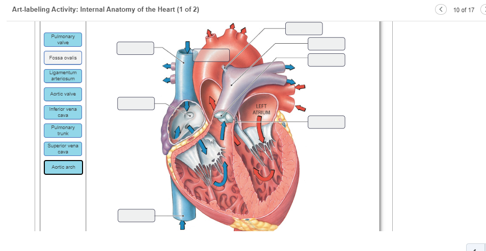


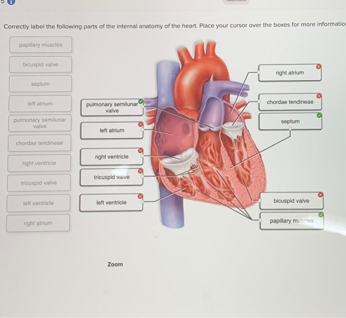



:max_bytes(150000):strip_icc()/VWH-MiraNorian-HeartElectricalSystem-Slide1-ba5b4bf3fdd2402da02cb0a48acdb8fe.jpg)










Post a Comment for "45 label the internal anatomy of the heart."