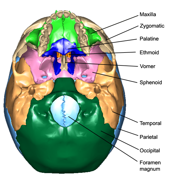
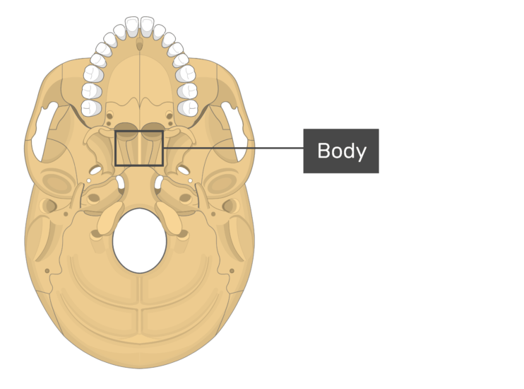
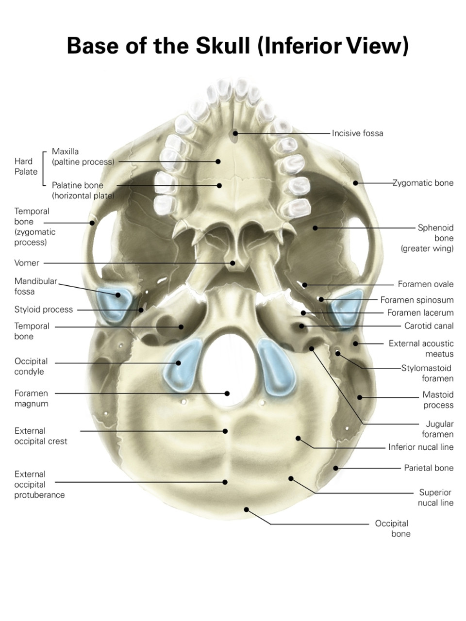
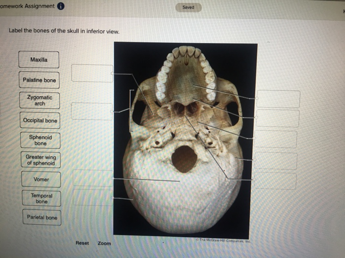
45 label the inferior bones and features of the skull
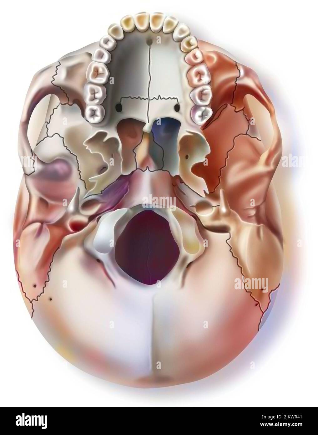
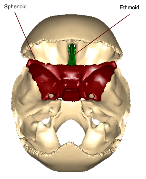
Lab 14: Figure 14.3 Inferior Bones of the Skull Diagram Start studying Lab 14: Figure 14.3 Inferior Bones of the Skull. Learn vocabulary, terms, and more with flashcards, games, and other study tools. Skull: Anatomy, structure, bones, quizzes | Kenhub Feb 8, 2023 · The skull base is the inferior portion of the neurocranium. Looking at it from the inside it can be subdivided into the anterior, middle and posterior cranial fossae. The skull base comprises parts of the frontal, ethmoid, sphenoid, occipital and temporal bones.
7.3 The Skull – Anatomy & Physiology Web26. Sept. 2019 · On the inferior aspect of the skull, each half of the sphenoid bone forms two thin, vertically oriented bony plates. These are …
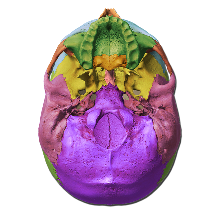
Label the inferior bones and features of the skull
Solved Label the bones and bone features shown on the - Chegg Label the bones and bone features shown on the inferior view of the skull by clicking and dragging the labels to the correct location Styloid process Zygomatic arch Mandibular fossa External acoustic meatus Mastold process rences Palatine bone Temporal bone Zygomatic bone Ootipal condylo Foramen magnum This problem has been solved! Figure 14.1 Label the anterior bones and features of the skull. (If the ... 1 9. 20. (bone). Page 2. Figure 14.3 Label the inferior bones and features ofthe skull. (bone) o '. 8. I. 1 0. (bone). Cranial Bones: Function and Anatomy, Diagram, Conditions ... - Healthline Anatomy and function. There are eight cranial bones, each with a unique shape: Frontal bone. This is the flat bone that makes up your forehead. It also forms the upper portion of your eye sockets ...
Label the inferior bones and features of the skull. The Skull Bones Anatomy - Inferior View | GetBodySmart Let's start with taking a look at the cranial and facial bones from an anterior view before we dive into their markings from an inferior perspective. Facial Bones: Zygomatic bone ( os zygomaticum ). Maxilla bone ( os maxilla ). Palatine bone ( os palatinum ). Learn skull anatomy faster with these interactive skull bones quizzes and worksheets. View of the Skull - Inferior - SmartDraw Create healthcare diagrams like this example called View of the Skull - Inferior in minutes with SmartDraw. SmartDraw includes 1000s of professional healthcare and anatomy chart templates that you can modify and make your own. 33/37 EXAMPLES. EDIT THIS EXAMPLE. Lab 14: Figure 14.3 Inferior Bones of the Skull Diagram Lab 14: Figure 14.3 Inferior Bones of the Skull 5.0 (3 reviews) + − Learn Test Match Created by THagge Teacher Terms in this set (15) Zygomatic Arch ... Vomer Bone ... Temporal Bone ... Maxilla ... Palatine Process of Maxilla ... Zygomatic Bone ... Palatine Bone ... Sphenoid Bone ... Styloid Process ... Exteral Acoustic Meatus ... Occipital Process Bones of the Skull - Structure - Fractures - TeachMeAnatomy The cranium (also known as the neurocranium) is formed by the superior aspect of the skull. It encloses and protects the brain, meninges, and cerebral vasculature. Anatomically, the cranium can be subdivided into a roof and a base: Cranial roof - comprised of the frontal, occipital and two parietal bones. It is also known as the calvarium.
Solved Label the bones and anatomical features of the - Chegg Label the bones and anatomical features of the inferior view of the skull, 16 Parietal hone Zygomatic arch Mastoid notch eBook Condylar canal Ext. Acoustic Meatus References The temporalis muscle passos modial to this structure. Sphenoid bone Mastoid process Occiptial Condyle This problem has been solved! Inferior view of the base of the skull: Anatomy | Kenhub Web20. Dez. 2022 · The sphenoid bone sits within the centre of the skull base like a wedge. This bone articulates with the vomer inferiorly, and the greater wings extend laterally to form part of the anterior pterion joint. From the … The Skull: Names of Bones in the Head, with Anatomy, & Labeled Diagram The zygomatic , lacrimal , palatine , vomer, nasal bones, and the inferior nasal concha form the rest of the orbits. Sutures of the Skull Sutures are a unique type of fibrous joint that connect the skull bones. These joints allow for movement during infancy, so the brain and skull can grow. Solved Label the bones and bone features shown on the WebQuestion: Label the bones and bone features shown on the inferior view of the skull by clicking and dragging the labels to the correct location Styloid process Zygomatic …
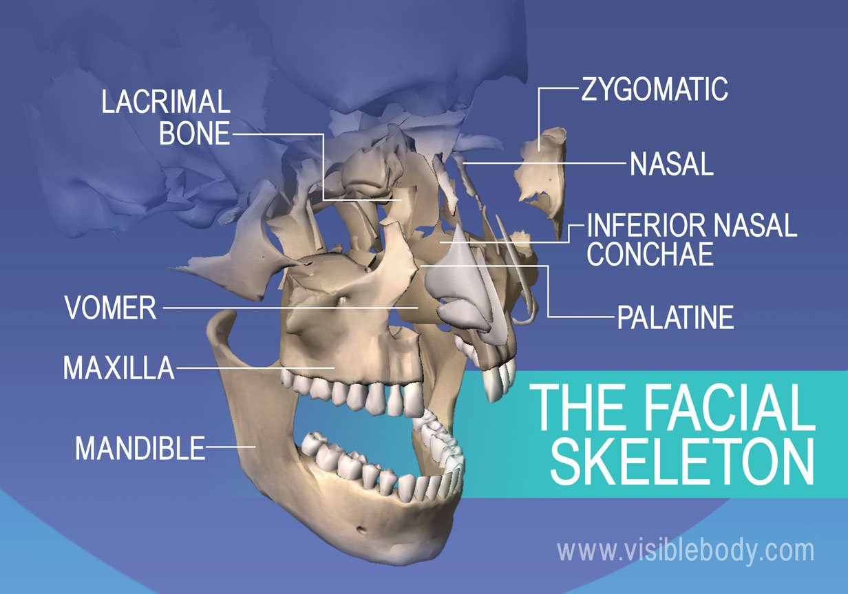
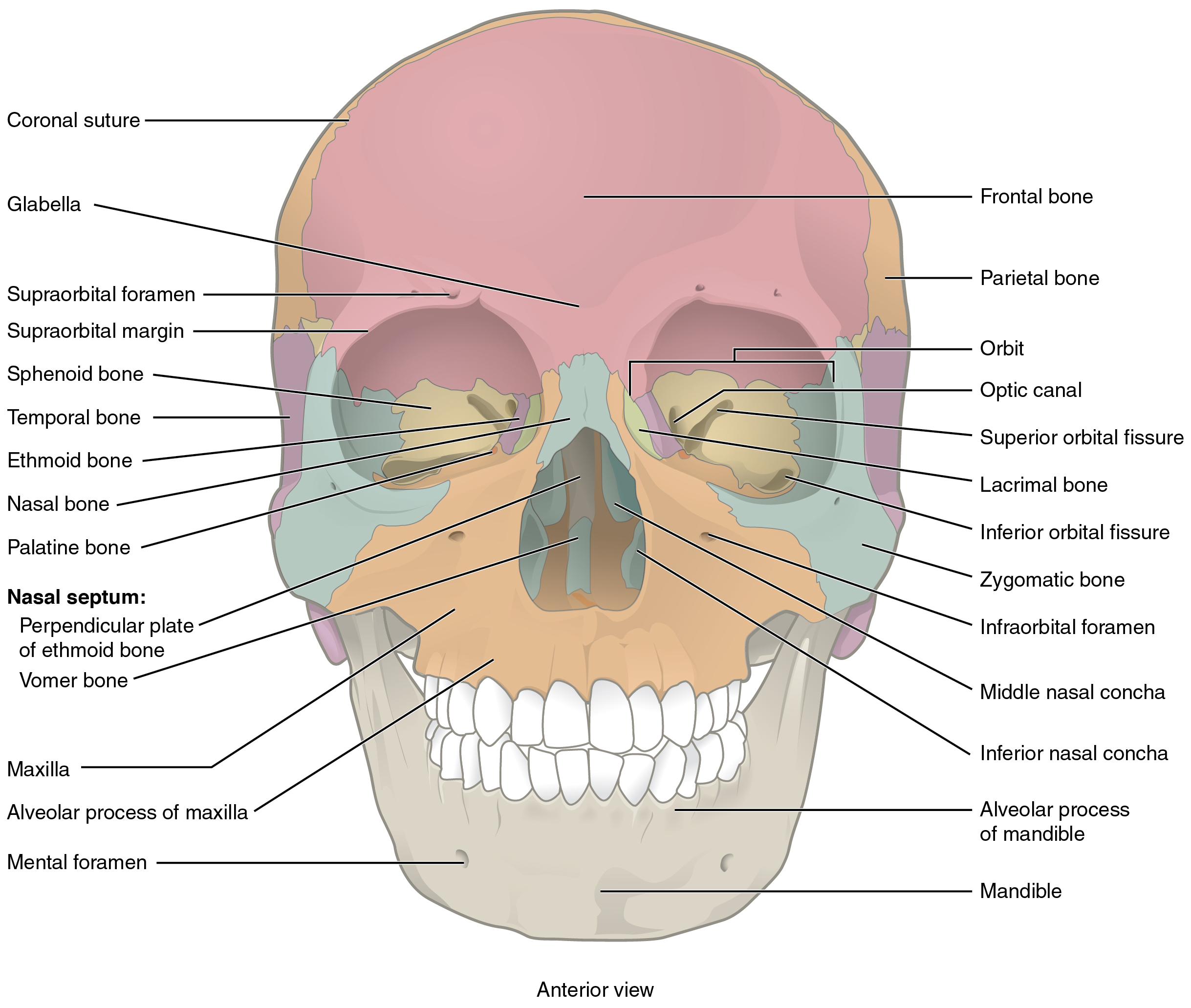
Lab 14: Figure 14.12 Inferior View of the Skull Diagram | Quizlet WebSets found in the same folder. Lab 14: Figure 14.11 Lateral View of the Skull. 14 terms Diagram. THagge Teacher. Lab 14: Figure 14.10 Anterior Features of the…. 9 terms … Skull Labeling - Inferior view Flashcards | Quizlet WebSkull Labeling - Inferior view 5.0 (1 review) zygomatic bone Click the card to flip 👆 Click the card to flip 👆 1 / 15 Flashcards Learn Test Match Created by ryanjmartin98 Terms in this … OpenStax AnatPhys fig.7.8 - Superior-Inferior View of Skull Base English labels. From OpenStax book 'Anatomy and Physiology', fig. 7.8. Anatomical structures in item: Bone. Cranium. Arcus zygomaticus. Os sphenoidale. 7.3 The Skull - Anatomy & Physiology The skull is the skeletal structure of the head that supports the face and protects the brain. It is subdivided into the facial bones and the cranium, or cranial vault (Figure 7.3.1).The facial bones underlie the facial structures, form the nasal cavity, enclose the eyeballs, and support the teeth of the upper and lower jaws.
The Skull: Names of Bones in the Head, with Anatomy, & Labeled … WebThe skull is one of the most vital bony structures of the human body, as it houses and protects the most important organs, including the brain. There are 29 bones (including …
Skull Labeling - Inferior view Flashcards | Quizlet Anatomy Skull Labeling - Inferior view 5.0 (1 review) zygomatic bone Click the card to flip 👆 Click the card to flip 👆 1 / 15 Flashcards Learn Test Match Created by ryanjmartin98 Terms in this set (15) zygomatic bone sphenoid bone vomer zygomatic process of temporal bone styloid process mastoid process occipital condyle temporal bone C.
Skull Anatomy | With Labels: Updated Version - YouTube Official Ninja Nerd Website: Nerds!In this lecture Professor Zach Murphy will present on the anatomy of the skull through the use ...
3. Lab 14: Figure 14.3 Inferior Bones of the Skull Diagram | Quizlet Start studying 3. Lab 14: Figure 14.3 Inferior Bones of the Skull. Learn vocabulary, terms, and more with flashcards, games, and other study tools.
Lab 14: Figure 14.3 Inferior Bones of the Skull Diagram WebLab 14: Figure 14.3 Inferior Bones of the Skull 5.0 (2 reviews) Learn Test Match Created by allisonrosentreter Terms in this set (15) Term Zygomatic Arch Location Term Vomer …
8.2.3: Markings of the Cranium - Biology LibreTexts WebMarkings of the Cranium Attributions (All Skull Sections) Markings of the Cranium Recall from Chapter 7: Introduction to the Skeletal System, that bones have markings including holes, passageways, basins, and …
Inferior view of the base of the skull: Anatomy | Kenhub Dec 20, 2022 · On the outer surface of the occipital bone is the inferior nuchal line. This is a small ridge of bone that projects from the posterior portion of the occipital bone. It gives attachment to a number of neck muscles including the rectus capitis posterior major and minor as well as the obliquus capitis superior.
The Skull | Anatomy and Physiology I - Lumen Learning On the inferior aspect of the skull, each half of the sphenoid bone forms two thin, vertically oriented bony plates. These are the medial pterygoid plate and lateral pterygoid plate (pterygoid = "wing-shaped"). The right and left medial pterygoid plates form the posterior, lateral walls of the nasal cavity.
Lab 14: Figure 14.3 Inferior Bones of the Skull Diagram | Quizlet WebLab 14: Figure 14.3 Inferior Bones of the Skull 5.0 (3 reviews) + − Learn Test Match Created by THagge Teacher Terms in this set (15) Zygomatic Arch ... Vomer Bone ...
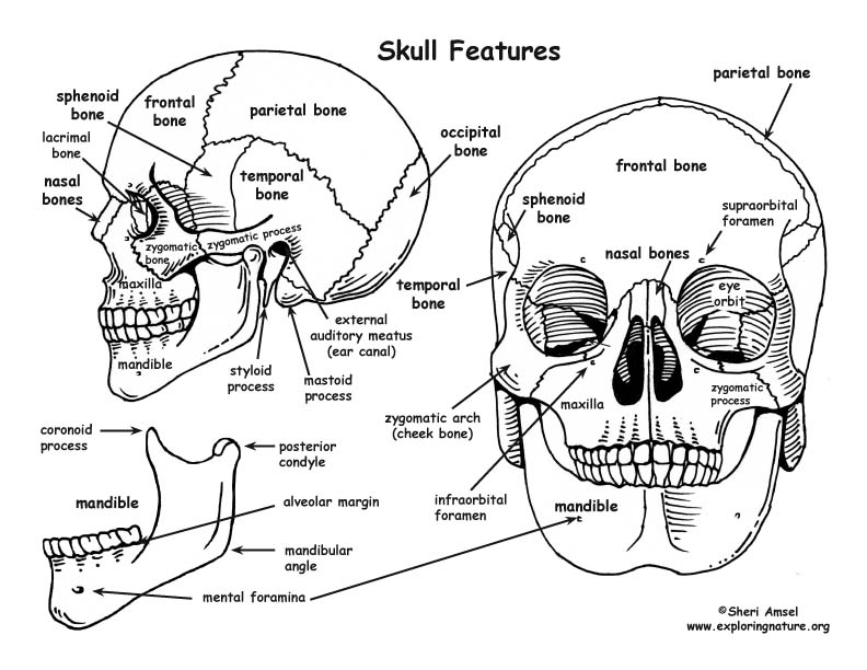
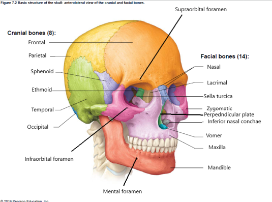
7.2 The Skull - Anatomy and Physiology 2e | OpenStax Identify the bones and structures that form the nasal septum and nasal conchae, and locate the hyoid bone. Identify the bony openings of the skull. The cranium (skull) is the skeletal structure of the head that supports the face and protects the brain. It is subdivided into the facial bones and the brain case, or cranial vault ( Figure 7.3 ).
Skull | Definition, Anatomy, & Function | Britannica skull, skeletal framework of the head of vertebrates, composed of bones or cartilage, which form a unit that protects the brain and some sense organs. The upper jaw, but not the lower, is part of the skull. The human cranium, the part that contains the brain, is globular and relatively large in comparison with the face. In most other animals the facial portion of the skull, including the upper ...
skull labeling.pdf 14. (bone). 15. (bone). 16. (bone). 17. (bone). 18. (bone). 19. (bone). 20. (bone). 21. Page 2. } Figure 12.3 Label the inferior bones and features of the skull ...
Cranial Bones: Function and Anatomy, Diagram, Conditions ... - Healthline Anatomy and function. There are eight cranial bones, each with a unique shape: Frontal bone. This is the flat bone that makes up your forehead. It also forms the upper portion of your eye sockets ...
Figure 14.1 Label the anterior bones and features of the skull. (If the ... 1 9. 20. (bone). Page 2. Figure 14.3 Label the inferior bones and features ofthe skull. (bone) o '. 8. I. 1 0. (bone).
Solved Label the bones and bone features shown on the - Chegg Label the bones and bone features shown on the inferior view of the skull by clicking and dragging the labels to the correct location Styloid process Zygomatic arch Mandibular fossa External acoustic meatus Mastold process rences Palatine bone Temporal bone Zygomatic bone Ootipal condylo Foramen magnum This problem has been solved!

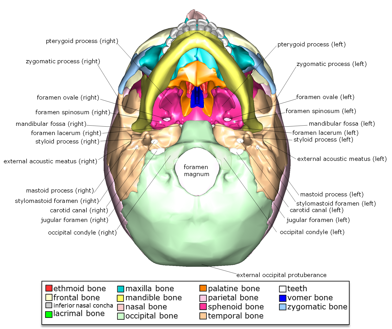


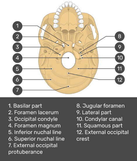

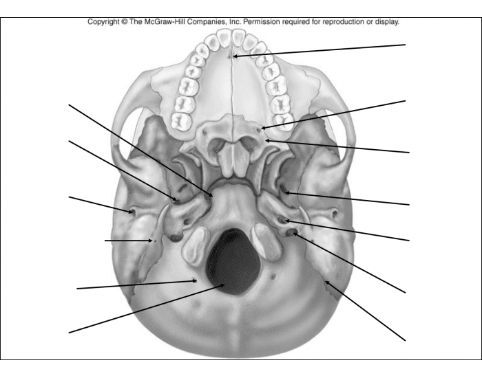




:watermark(/images/watermark_5000_10percent.png,0,0,0):watermark(/images/logo_url.png,-10,-10,0):format(jpeg)/images/overview_image/361/VamnYnBlLvYkS8hAb2S2FQ_inferior-base-of-the-skull-landmarks_english.jpg)










Post a Comment for "45 label the inferior bones and features of the skull"