39 correctly label the structures associated with unmyelinated nerve fibers in the pns
Question: CORRECTLY LABEL THE STRUCTURES ASSOCIATED WITHUNMYELINATED ... Correctly label the structures associated with unmyelinated nerve fibers in the PNS Schwann cel Unmyelinated nerve nbers Basal lamina ?? Post navigation Question: Identify which type of microscope would be most appropriate toobserve each of the following samp [Unmyelinated nerve fibers] - PubMed [Unmyelinated nerve fibers] [Article in Russian] Author O S Sotnikov 1 Affiliation 1 Laboratory of Neuron Functional Morphology and Physiology, RAS I.P. Pavlov Institute of Physiology, St. Petersburg. PMID: 12891789 Abstract The paper presents a critical review of various current concepts of the structure and kinetics of unmyelinated nerve fiber.
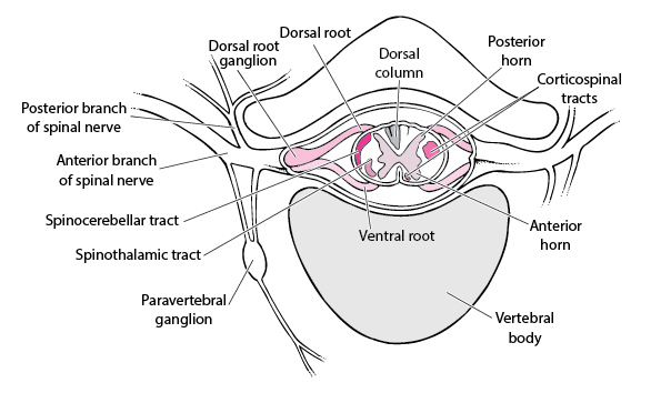
Unmyelinated Nerve Fiber - an overview | ScienceDirect Topics Nerve impulses generated at the nociceptive receptor system are delivered into the spinal cord by small myelinated and unmyelinated nerve fibres (5 µ m or less in diameter), that mainly belong to the Ad and C groups of afferent nerve fibres ( Fig. 1.1 ). Their cell bodies are located in the dorsal root ganglia of the spinal nerves.
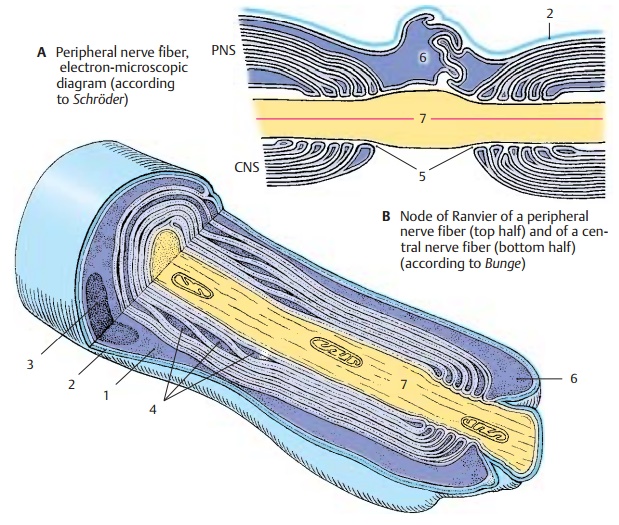
Correctly label the structures associated with unmyelinated nerve fibers in the pns
Question: CORRECTLY LABEL THE STRUCTURES ASSOCIATED WITH UNMYELINATED ... UNMYELINATED FIBERS IN THE PNS. Correctly label the structures associated with unmyelinated nerve fibers in the PNS Schwann cel Unmyelinated nerve nbers Basal lamina 閰咇 [supanova_question] Question: Ependymal cell Oligodendrocyte Neurons MicrogliaMyelinated axon Capillary Astrocyte Prex 4 of 50 … college essay help free ... What is true about unmyelinated nerve fiber? - Byju's Unmyelinated nerve fibres are those which are enclosed by Schwann cells however these Schwann cells do not form myelin sheath around the axon. Unmyelinated nerve fibres are found in autonomous and somatic neural systems. Hence option b is correct in respect to unmyelinated nerve fibres. Suggest Corrections. (PDF) Unmyelinated nerve fibers in the human dental pulp express ... Unmyelinated nerve fibers in the human dental pulp express markers for myelinated fibers and show sodium channel accumulations . × Close Log In. Log in with Facebook Log in with Google. or. Email. Password. Remember me on this computer. or reset password. Enter the email address you signed up with and we'll email you a reset link. ...
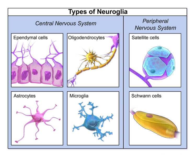
Correctly label the structures associated with unmyelinated nerve fibers in the pns. Peripheral nerves: Histology and clinical aspects | Kenhub Unmyelinated fibers (histological slide) Skeletal muscles cells also have modified synaptic interactions with telodendria known as myoneural junctions (motor end plates). A single motor neuron may innervate several to several hundred muscle cells; collectively the unit is referred to as a motor end plate. Unmyelinated Nerve Fiber - an overview | ScienceDirect Topics (b) The afferent fibers were identified as unmyelinated by electrical stimulation of the neuroma with single pulses. The signal indicated by the red dot was recorded from the same afferent fiber as in (c) and (d). (c) Record from a single unmyelinated afferent fiber. AHCDW8Notes10.pdf - 10. Award: 10.00 points Problems?... Correctly label the structures associated with unmyelinated nerve fibers in the PNS. Explanation:In an unmyelinated nerve fiber multiple unmyelinated fibers are enclosed in channels in the surface of a single Schwann cell. Nerves: The Histology Guide - University of Leeds The peripheral nervous system (PNS) consists of nerves, which are bundles of nerve fibres, ganglia, which contain not only nerve fibres but also neuronal cell bodies (perikarya) and nerve endings. In the PNS, nerves contain, in addition to fibres, specialised supporting cells, called Schwann cells. What is their function?
12 Difference Between Myelinated And Unmyelinated Nerve Fibers Myelinated Nerve Fibers are nerve fibers that are insulated by a myelin sheath. Unmyelinated nerve fibers are nerve fibers that do not have a myelin sheath. The nerve fibers with long axons are myelinated. The short axon nerve fibers are unmyelinated. The axis cylinder of the myelinated nerve fibres has two sheaths. Structure of the myelinated nerve fiber | Learn Science at Scitable The nerve tract is filled with ring-shaped structures, indicating a transverse section of myelinated nerve fibers. (D) Electron microscopy of a myelinated axon in a mouse optic nerve at... Unmyelinated nerve fibres enclosed by Schwann cells are commonly found in? The myelinated nerve fibres are enveloped with Schwann cells, it form a myelin sheath around the axon. Unmyelinated nerve fibres is enveloped by a Schwann cell that doesn't form a myelin sheath around the axon, and found in autonomous and the somatic neural systems. Sympathetic Nervous system is part of Autonomous Nervous system. Which of the following is true about myelinated axons? a. Propagates ... Which of the following structures IS NOT common to all nerve cells? A. cell body B. axons C. dendrite D. Schwann cells; The white matter of the spinal cord contains [{Blank}]. a. myelinated nerve fibers only. b. myelinated and unmyelinated nerve fibers. c. unmyelinated nerve fiber only. d. soma that have both myelinated and unmyelinated nerve ...
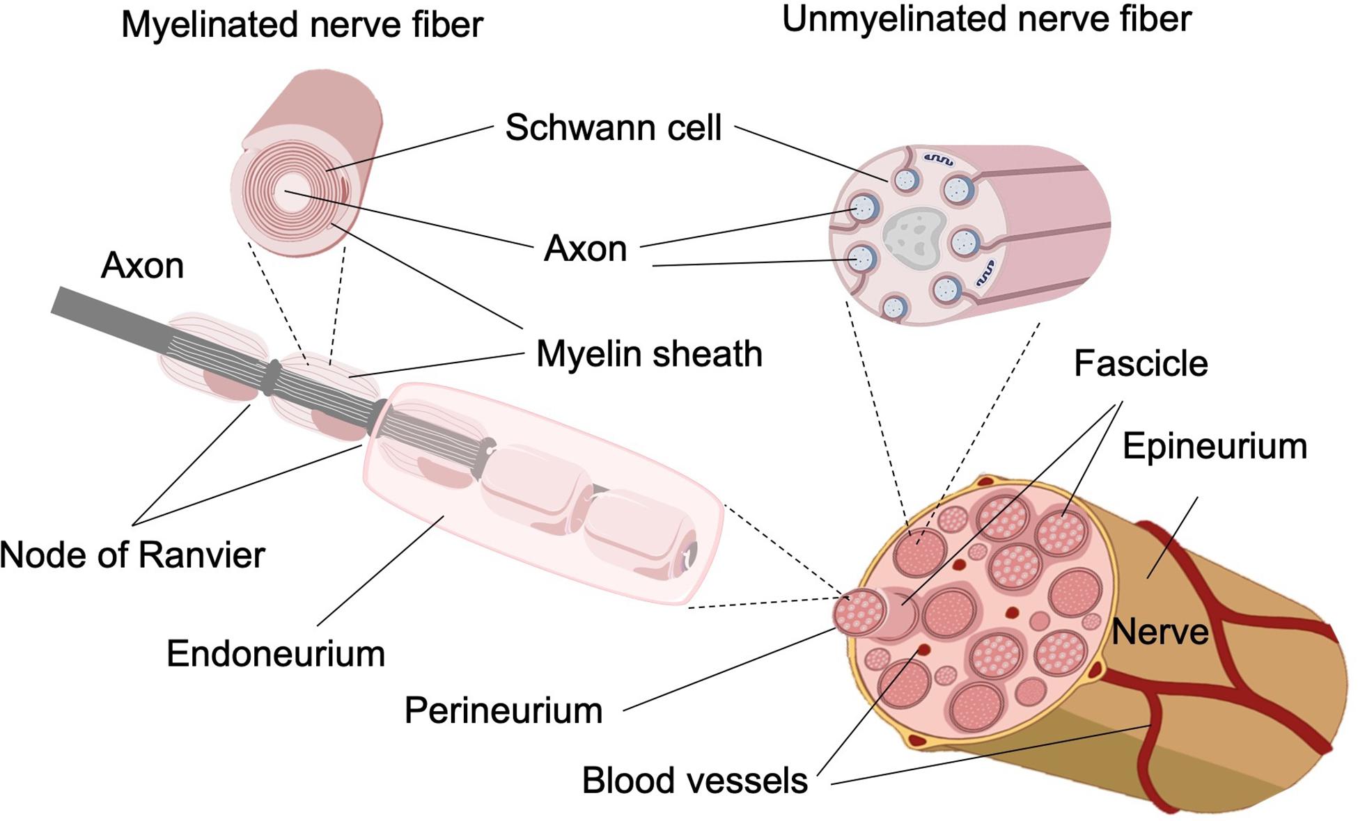
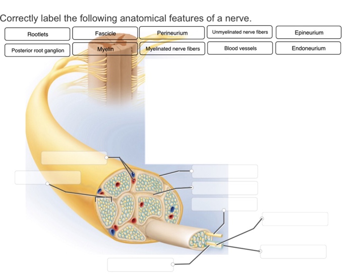
Nerve Fibers, Unmyelinated - MeSH - NCBI A class of nerve fibers as defined by their nerve sheath arrangement. The AXONS of the unmyelinated nerve fibers are small in diameter and usually several are surrounded by a single MYELIN SHEATH. They conduct low-velocity impulses, and represent the majority of peripheral sensory and autonomic fibers, but are also found in the BRAIN and SPINAL ... Ch.6: Nervous System - PNS - Histology - Nervous PNS A. PNS: Nerve ... Advanced Anatomy & Physiology for Health Professions (NUR 4904) Pharmacology (RNSG 1301) ... Histology - Nervous PNS A. PNS: Nerve Fibers a. Nerve ibers are axons sheathed by Schwann Cells (nerve iber is NOT a synonym for neuron cell) i. ... Slower transmission for unmyelinated nerve ibers/axons because not saltatory 1. Will have axons with ... The Peripheral Nervous System | SEER Training Each bundle of nerve fibers is called a fasciculus and is surrounded by a layer of connective tissue called the perineurium. Within the fasciculus, each individual nerve fiber, with its myelin and neurilemma, is surrounded by connective tissue called the endoneurium. A nerve may also have blood vessels enclosed in its connective tissue wrappings. Solved Correctly label the structures associated with - Chegg Question: Correctly label the structures associated with unmyelinated axons in the PNS Unmyelinated axons ID Neurolemmocyte Neurolemmocyte nucleus Show transcribed image text Expert Answer 100% (4 ratings) The uppermost structure is unmyelinated axons. T … View the full answer Transcribed image text:
51139882-C1CB-4A6C-ABC6-DB03F69453B6.jpeg - Correctly label... Correctly label the structures associated with unmyelinated nerve fibers in the PNS Study Resources Main Menu by School by Literature Title by Subject Textbook SolutionsExpert TutorsEarn Main Menu Earn Free Access Upload Documents Refer Your Friends Earn Money Become a Tutor Apply for Scholarship For Educators Log in
Solved CORRECTLY LABEL THE STRUCTURES ASSOCIATED WITH - Chegg CORRECTLY LABEL THE STRUCTURES ASSOCIATED WITH UNMYELINATED FIBERS IN THE PNS. Show transcribed image text Expert Answer 100% (22 ratings) Answer: Correctly … View the full answer Transcribed image text: Correctly label the structures associated with unmyelinated nerve fibers in the PNS Schwann cel Unmyelinated nerve nbers Basal lamina 閰咇
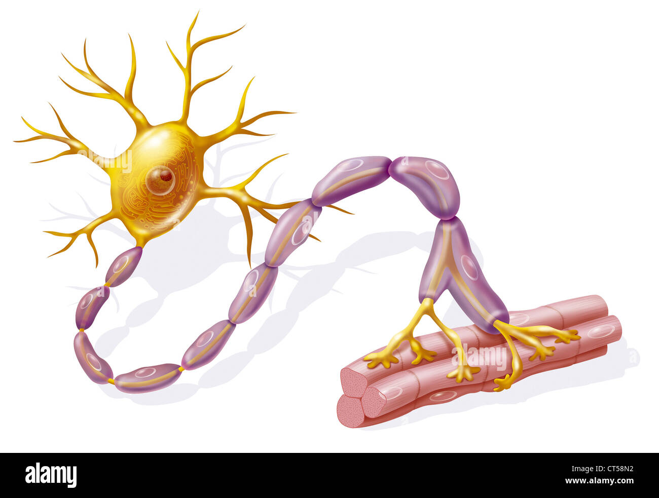
Unmyelinated Nerve Fibers - Physiology - AmeriCorps Health Many nerve fibers in the CNS and PNS are unmyelinated. In the PNS, however, even the unmyelinated fibers are enveloped in Schwann cells. In this case, one Schwann cell harbors from 1 to 12 small nerve fibers in grooves in its surface.
Chapter 12 QS Anatomy (Nervous System) Flashcards | Quizlet Label the structures of a nerve. Label the figure with the items provided. bipolar neuron, anaxonic neuron, unipolar neuron, multipolar neuron If all the sodium leakage channels were removed from the cell membrane of a neuron, the membrane potential would be about -90 millivolts. Label the features of a myelinated axon.
Difference Between Myelinated and Unmyelinated Nerve Fibers The unmyelinated nerve fibers are gray in color. Most of their axons are short. The peripheral postganglionic autonomic fibers are a type of unmyelinated nerve fibers. The C fibers of the skin, muscles, and viscera are also unmyelinated fibers. The olfactory nerves are also unmyelinated. Figure 2: Myelinated and Unmyelinated Nerve Fibers
12 Difference Between Myelinated And Unmyelinated Neurons (Nerve Fiber ... Unmyelinated nerve fibers are nerve fibers that do not have a myelin sheath. The short axon nerve fibers are unmyelinated. The axis cylinder of unmyelinated nerve fibres has only one sheath. Unmyelinated nerve fibres do not show notes and internodes. The nerve fibers appear gray in color.
Peripheral Nervous System | histology The CNS consists of the brain and the spinal cord, while the PNS is composed of nerves and groups of nerve cells (neurons), called ganglia. The nerves of the PNS carry sensory (afferent) inputs to the CNS and motor (efferent) output from the CNS to the skeletal and cardiac muscles and the smooth muscles of blood vessels, organs and glands.
Duke Histology - Nerve Tissue In the case of unmyelinated axons, the unmyelinated fiber shares each Schwann cell with several other unmyelinated axons. Nerve = a bundle or bundles of nerve fibers. Ganglion = clusters of neuronal cell bodies in the peripheral nervous systems, as well as associated glial cells and axons. Therefore, ganglia can be distinguished from peripheral ...
Lab 1 Homework BIOL 320 Flashcards | Quizlet Correctly label the structures associated with unmyelinated nerve fibers in the PNS. Indicate the specified region of the diencephalon. Thalamus Determine which general anatomic feature of the brain is illustrated in the figure. Sulcus Determine which specific tract is depicted in the figure.
Neuroanatomy, Unmyelinated Nerve Fibers - PubMed C-type fibers are unmyelinated fibers that form one of these groups, and they are involved in the afferent transfer of temperature, burning pain, and itch from the periphery to synapse, principally in lamina I and II of the dorsal spinal horn. Copyright © 2022, StatPearls Publishing LLC. Sections Introduction Structure and Function
(PDF) Unmyelinated nerve fibers in the human dental pulp express ... Unmyelinated nerve fibers in the human dental pulp express markers for myelinated fibers and show sodium channel accumulations . × Close Log In. Log in with Facebook Log in with Google. or. Email. Password. Remember me on this computer. or reset password. Enter the email address you signed up with and we'll email you a reset link. ...
What is true about unmyelinated nerve fiber? - Byju's Unmyelinated nerve fibres are those which are enclosed by Schwann cells however these Schwann cells do not form myelin sheath around the axon. Unmyelinated nerve fibres are found in autonomous and somatic neural systems. Hence option b is correct in respect to unmyelinated nerve fibres. Suggest Corrections.
Question: CORRECTLY LABEL THE STRUCTURES ASSOCIATED WITH UNMYELINATED ... UNMYELINATED FIBERS IN THE PNS. Correctly label the structures associated with unmyelinated nerve fibers in the PNS Schwann cel Unmyelinated nerve nbers Basal lamina 閰咇 [supanova_question] Question: Ependymal cell Oligodendrocyte Neurons MicrogliaMyelinated axon Capillary Astrocyte Prex 4 of 50 … college essay help free ...
















Post a Comment for "39 correctly label the structures associated with unmyelinated nerve fibers in the pns"