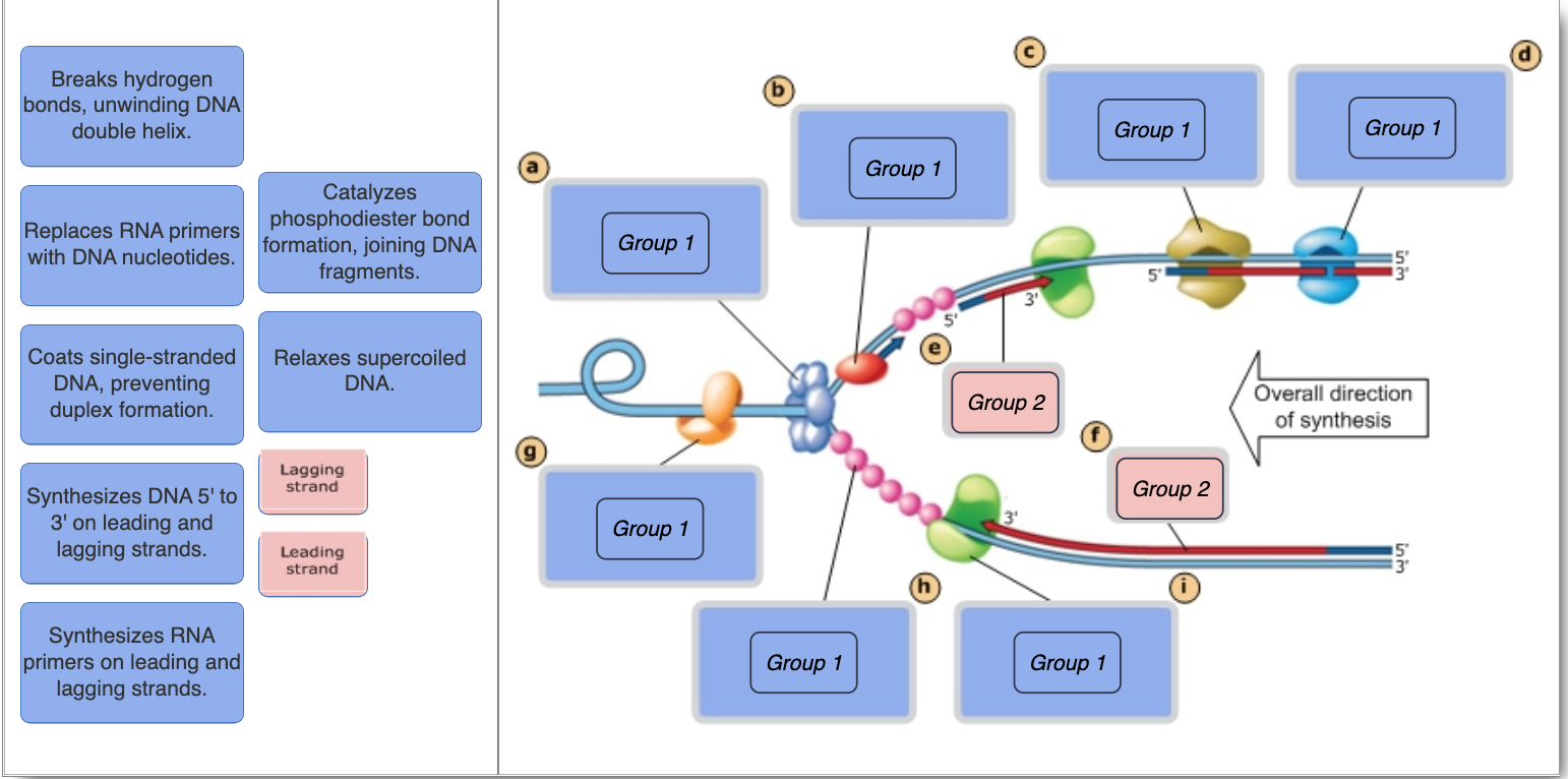
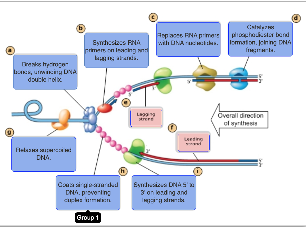
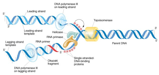
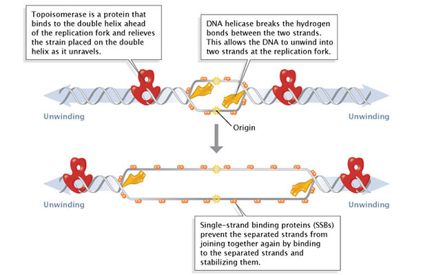
42 the diagram below shows a bacterial replication fork and its principal proteins. drag the labels to their appropriate locations in the diagram to describe the name or function of each structure
Mastering Biology Chp. 13 HW - Subjecto.com Mastering Biology Chp. 13 HW Home » Flashcards » Mastering Biology Chp. 13 HW Flashcards Total word count: 4468 Pages: 16 Get Now Calculate the Price Deadline Paper type Pages - - 275 words Check Price Looking for Expert Opinion? Let us have a look at your work and suggest how to improve it! Get a Consultant « Previous Flashcard Next Flashcard » Chapter 11 Flashcards | Quizlet The diagram below shows a bacterial replication fork and its principal proteins. Drag the labels to their appropriate locations in the diagram to describe the name or function of each structure. Use pink labels for the pink targets and blue labels for the blue targets. A) Breaks hydrogen bonds, unwinding DNA double helix.
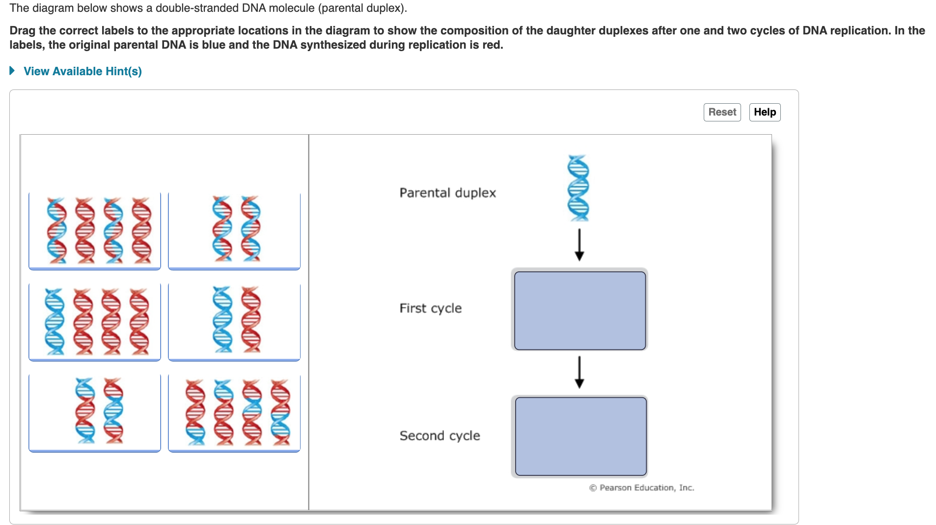
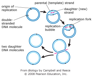
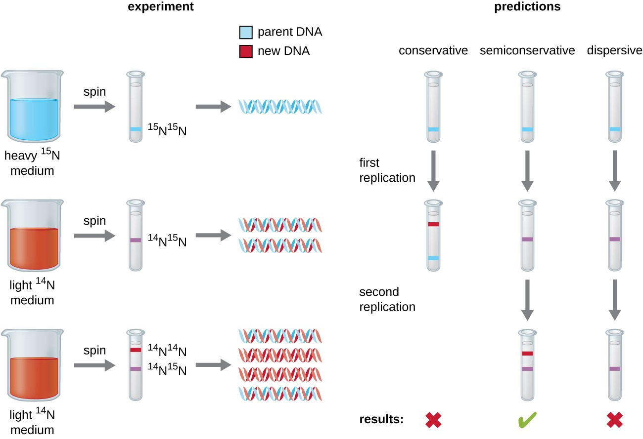
Question 1.pdf - 2/17/2020 MasteringBiology: Ch 15 HW... 2/17/2020 MasteringBiology: Ch 15 HW 1/1 Part A - The mechanism of DNA replication The diagram below shows a double-stranded DNA molecule (parental DNA). Drag the correct labels to the appropriate locations in the diagram to show the composition of the daughter DNA molecules after one and two cycles of DNA replication. In the labels, the original parental DNA is blue and the DNA synthesized ...

The diagram below shows a bacterial replication fork and its principal proteins. drag the labels to their appropriate locations in the diagram to describe the name or function of each structure
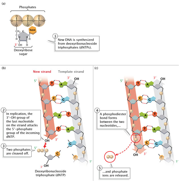
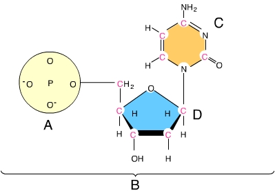
Mastering Biology Chapter 13 Flashcards | Quizlet The first, marked A, contains a phosphorus atom with four oxygen atoms. Two oxygen atoms have negative charge. Letter B marks a pentagon with an O at its top vertex and a CH2OH attached above its left vertex. One can numerate carbon atoms clockwise starting from next to O. Then OH group is attached below to third carbon atom. the diagram below shows a bacterial replication fork and its principal ... the diagram below shows a bacterial replication fork and its principal proteins. Replication chromosome integrity genome If you are looking for The Diagram Below Shows A Bacterial Replication Fork And Its Principal you've visit to the right place. The diagram below shows a bacterial replication fork and its principal ... The diagram below shows a bacterial replication fork and its principal proteins. Drag the labels to their appropriate locations in the diagram to describe the name or function of each structure. Use pink labels for the pink targets and blue labels for the blue targets. The diagram below shows a bacterial replication fork and its principal proteins.
The diagram below shows a bacterial replication fork and its principal proteins. drag the labels to their appropriate locations in the diagram to describe the name or function of each structure. Ch 15 HW.pdf - Ch15HW Due:9:00pmonSunday,February19,2017... - Course Hero Drag the correct labels to the appropriate locations in the diagram to show the composition of the daughter DNA molecules after one and two cycles of DNA replication. In the labels, the original parental DNA is blue and the DNA synthesized during replication is red. You did not open hints for this part. Part B Processes occurring at a bacterial replication fork The diagram ... Match each protein with its function in bacterial DNA replication. ANSWER: HelpReset Reset Help 1. Primase synthesizes short sequences of RNA required for DNA replication. 2. Helicase breaks the hydrogen bonds that hold the parental DNA strands together. 3. Single-strand binding protein coats the separated DNA strands. 4. Drag The Labels To Their Appropriate Locations In This Diagram The diagram below shows a bacterial replication fork and its principal proteins. Drag The Labels To Their Appropriate Locations In The Diagram To Describe The Name Or Function Of. A network diagram can be either physical or logical. Drag the labels to their appropriate locations on the diagram below. Then use the labels of group 2 to identify the. Mastering Biology Chp. 13 HW Flashcards | Quizlet In DNA replication in bacteria, the enzyme DNA polymerase III (abbreviated DNA pol III) adds nucleotides to a template strand of DNA. But DNA pol III cannot start a new strand from scratch. Instead, a primer must pair with the template strand, and DNA pol III then adds nucleotides to the primer, complementary to the template strand.
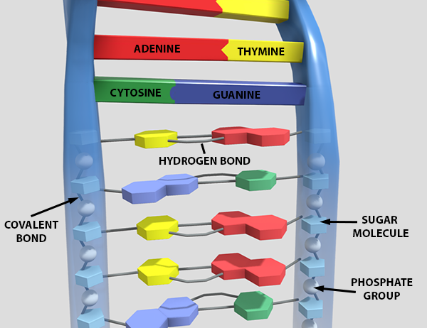
Drag the Labels to the Appropriate Locations in This Diagram. The diagram below shows a bacterial replication fork and its principal proteins.. Drag the labels to their appropriate locations on the diagram. Use only the blue labels for the blue targets and only the pink labels for the pink targets. Changes in cytosolic Ca 2 concentration link action potentials in the muscle cell to contraction of the ... biology chapter 16 Flashcards | Quizlet In DNA replication in bacteria, the enzyme DNA polymerase III (abbreviated DNA pol III) adds nucleotides to a template strand of DNA. But DNA pol III cannot start a new strand from scratch. Instead, a primer must pair with the template strand, and DNA pol III then adds nucleotides to the primer, complementary to the template strand. the diagram below shows a bacterial replication fork and its principal ... The diagram below shows a bacterial replication fork and its principal proteins. The diagram below shows a bacterial replication fork and its principal proteins. Drag the labels to their appropriate locations in the diagram to describe the name or function of each structure. Use pink labels for the pink targets and blue labels for the blue targets. Mastering Biology ch. 13 Flashcards | Quizlet Drag the labels to their appropriate locations on the diagram below. Targets of Group 1 can be used more than once. b. 3' end The DNA double helix is composed of two strands of DNA; each strand is a polymer of DNA nucleotides. Each nucleotide consists of a sugar, a phosphate group, and one of four nitrogenous bases.
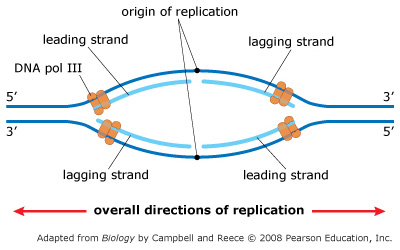
DNA replication and transcription Post-Lecture - Quizlet The Taq enzyme is a type of DNA polymerase that allows researchers to separate the DNA strands during the annealing step of the PCR cycle without destroying the polymerase. False How many DNA molecules would there be after four rounds of PCR if the initial reaction mixture contained two molecules? 32 Mastering Biology Chapter 16 - RHS Homework The diagram below shows a replication bubble with synthesis of the leading and lagging strands on both sides of the bubble. The parental DNA is shown in dark blue, the newly synthesized DNA is light blue, and the RNA primers associated with each strand are red. The origin of replication is indicated by the black dots on the parental strands. The diagram below shows a bacterial replication fork and its principal ... The diagram below shows a bacterial replication fork and its major proteins. Drag labels to their appropriate locations on the diagram to describe the name or function of each structure. Use pink tags for pink targets and blue tags for blue targets. Answer and. end f. leading to (a) breaks hydrogen bonds, unwinding the DNA double helix. intro to cell test five Flashcards | Quizlet The following eukaryotic structural gene contains two introns and three exons. The table below shows four possible mRNA products of this gene. Use the labels to explain what mutation (s) may have resulted in each mRNA. Drag the correct label to each location in the table. Labels may be used once, more than once, or not at all.
Solved The diagram below shows a bacterial replication fork - Chegg Question: The diagram below shows a bacterial replication fork and its principal proteins. Drag the labels to their appropriate locations in the diagram to describe the name or function of each structure. Use pink labels for the pink targets and blue labels for the blue targets.
The diagram below shows a bacterial replication fork and its principal ... Use pink labels for the pink targets and blue labels for the blue targets. The diagram below shows a bacterial replication fork and its principal proteins. Drag the labels to their appropriate locations in the diagram to describe the name or function of each structure. Use pink labels for the pink targets and blue labels for the blue targets.
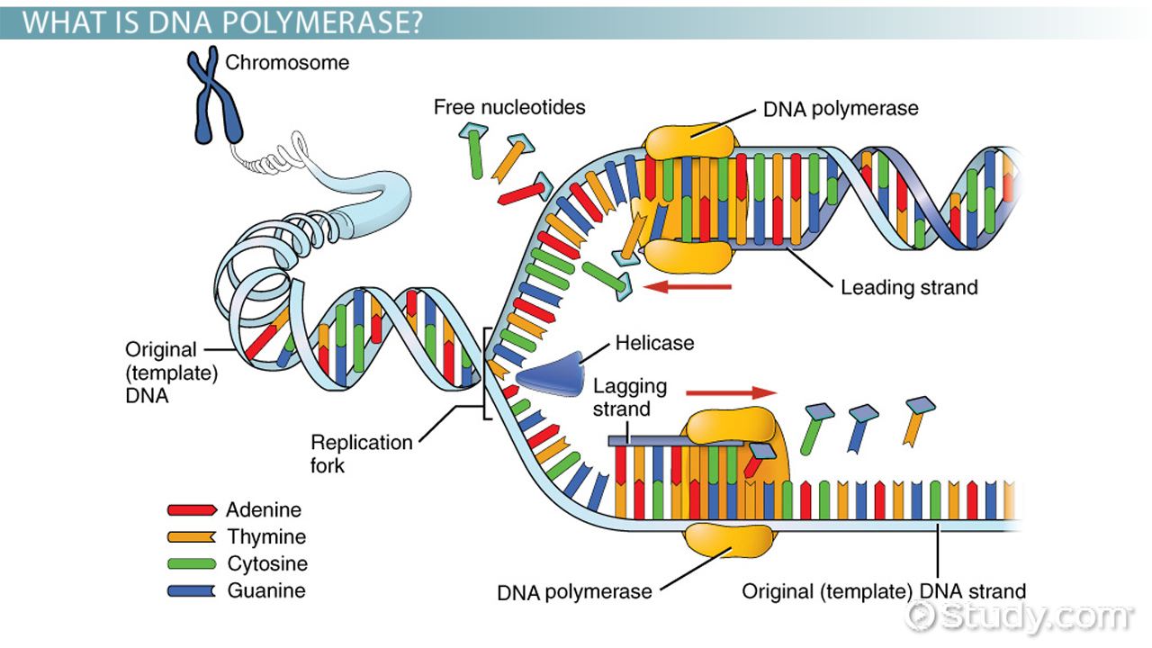
The diagram below shows a bacterial replication fork and its principal ... The labeled diagram of bacterial replication fork and its principal proteins. What does "replication fork" mean? The portion of DNA where the replication process is now underway is known as the replication fork. Its design is reminiscent of a fork. A multiprotein complex that accomplishes replication is located at the replication fork.
Replication Fork: Definition, Structure, Diagram, & Function Replication Fork. The replication fork is a structure which is formed during the process of DNA replication. It is activated by helicases, which helps in breaking the hydrogen bonds, and holds the two strands of the helix. The resulting structure has two branching's which is known as prongs, where each one is made up of single strand of DNA.
Drag each phrase to the appropriate bin depending on whether it ... Part B - Processes occurring at a bacterial replication fork The diagram below shows a bacterial replication fork and its principal proteins. Drag the labels to their appropriate locations in the diagram to describe the name or function of each structure. Hint 1. What are the functions of the proteins involved in bacterial DNA replication ...
The diagram below shows a bacterial replication fork and its principal ... The diagram below shows a bacterial replication fork and its principal proteins. Drag the labels to their appropriate locations in the diagram to describe the name or function of each structure. Use pink labels for the pink targets and blue labels for the blue targets. The diagram below shows a bacterial replication fork and its principal proteins.
the diagram below shows a bacterial replication fork and its principal ... the diagram below shows a bacterial replication fork and its principal proteins. Replication chromosome integrity genome If you are looking for The Diagram Below Shows A Bacterial Replication Fork And Its Principal you've visit to the right place.
Mastering Biology Chapter 13 Flashcards | Quizlet The first, marked A, contains a phosphorus atom with four oxygen atoms. Two oxygen atoms have negative charge. Letter B marks a pentagon with an O at its top vertex and a CH2OH attached above its left vertex. One can numerate carbon atoms clockwise starting from next to O. Then OH group is attached below to third carbon atom.


:max_bytes(150000):strip_icc()/DNA_replication_elongation2-e38fc92e8ad74586bfe0443d7490d3ce.jpg)













Post a Comment for "42 the diagram below shows a bacterial replication fork and its principal proteins. drag the labels to their appropriate locations in the diagram to describe the name or function of each structure"