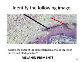
44 label the tissue and structures on this histology slide
Histology of the Endocrine System - MVCC Anatomy and Physiology Slides 1 and 2: Glandular and Nervous Tissues. Slide 1 - Submandibular Gland. Label A indicates two clusters of secretory cells. Label B points out two small ducts. ... Thyroid displays a unique structure that is lobular, however the center of the lobule is filled with a colloidal solution that is a precursor to the hormonal secretion of ... Epithelian tissue labeled slides Flashcards | Quizlet Brian Bich, Anatomy & Physiology I, Biology 1140, Lake Superior College, Slide images provided from Bish Terms in this set (32) Pseudostratified Ciliated Columnar Epithelial Tissues Pseudostratified Ciliated Columnar Epithelial Tissues Pseudostratified Ciliated Columnar Epithelial Tissues Pseudostratified Ciliated Columnar Epithelial Tissues
Learn histology faster With quizzes and flashcards | Kenhub With Kenhub's huge library of histology slides, of course! In our histology atlas, we clearly highlight a given structure on our slides. Comparing several of these slides next to one another is a great way to get a feel for how one tissue differs from another. Enter: our labeled and unlabeled histology tissue identification quiz worksheets.
Label the tissue and structures on this histology slide
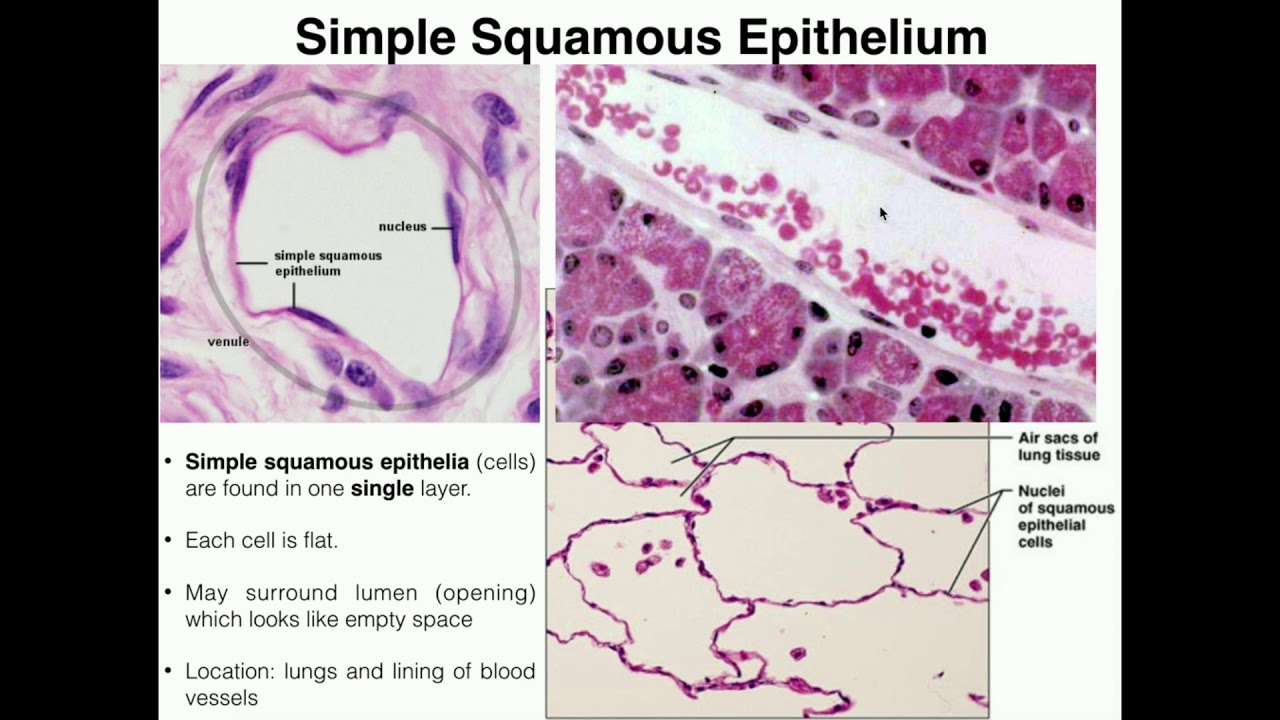
Skeletal Muscle Histology Slide Identification and Labeled Diagram ... From the skeletal muscle histology slide, you might identify the following important structures under the light microscope. Please try to find out these structures from the skeletal muscle slide labeled images. #1. Longitudinal section of skeletal muscle #2. Cross-section of skeletal muscle #3. Skeletal muscle fibers of the longitudinal section #3. Microscope Slides of Cells and Tissues | Histology Guide This virtual slide box contains 275 microscope slides for the learning histology. Fig 023 Types of Tissue Cells and Tissues Tissues are classified into four basic types: epithelium, connective tissue (includes cartilage, bone and blood), muscle, and nervous tissue. Chapter 1 The Cell Chapter 2 Epithelium Chapter 3 Connective Tissue Chapter 4 Muscle Histology guide: Definition and slides | Kenhub At a histological level, both the heart and blood vessels consist of three layers: Endothelial layer - epithelial tissue formed by simple squamous (endothelial) cells. In the heart, this layer is referred to as endocardium. Muscular layer - smooth muscle in the blood vessels, cardiac muscle (myocardium) in the heart.
Label the tissue and structures on this histology slide. › articles › s41592/021/01255-8SpaGCN: Integrating gene expression, spatial location and ... Oct 28, 2021 · a, Histology image of the tissue section and spatial domains detected by Louvain, stLearn, BayesSpace and SpaGCN. b , Spatial subdomains of the cortex region detected by Louvain, stLearn ... Activity 2 - Histology and Integument - slideshare.net 1. Activity 2: Histology and Integument Chapter 4 & 5 - Human Anatomy (4e) textbook Objectives: • Identify each tissue (26 tissues) in a histology photo or microscope slide. • Sketch each tissue in your lab manual. • Identify the features of the integument (skin) on a slide and/or model. 1 Compilation: Benjamin Sparks & Claudia Gonzales ... Histology of the Lymphatic System - SlideShare 2. Lymphatic System Consists of group of cells, tissues & organs that monitor body surfaces and internal fluid and react to potentially harmful substances. 3. Examples of immune response Reaction against microorganisms: bacteria, viruses, parasites Reaction against tumor cells Allergic reactions: Hay fever Autoimmune reaction: Arthritis Graft ... en.wikipedia.org › wiki › StainingStaining - Wikipedia Staining is a technique used to enhance contrast in samples, generally at the microscopic level. Stains and dyes are frequently used in histology (microscopic study of biological tissues) and in the medical fields of histopathology, hematology, and cytopathology that focus on the study and diagnoses of disease at a microscopic level.
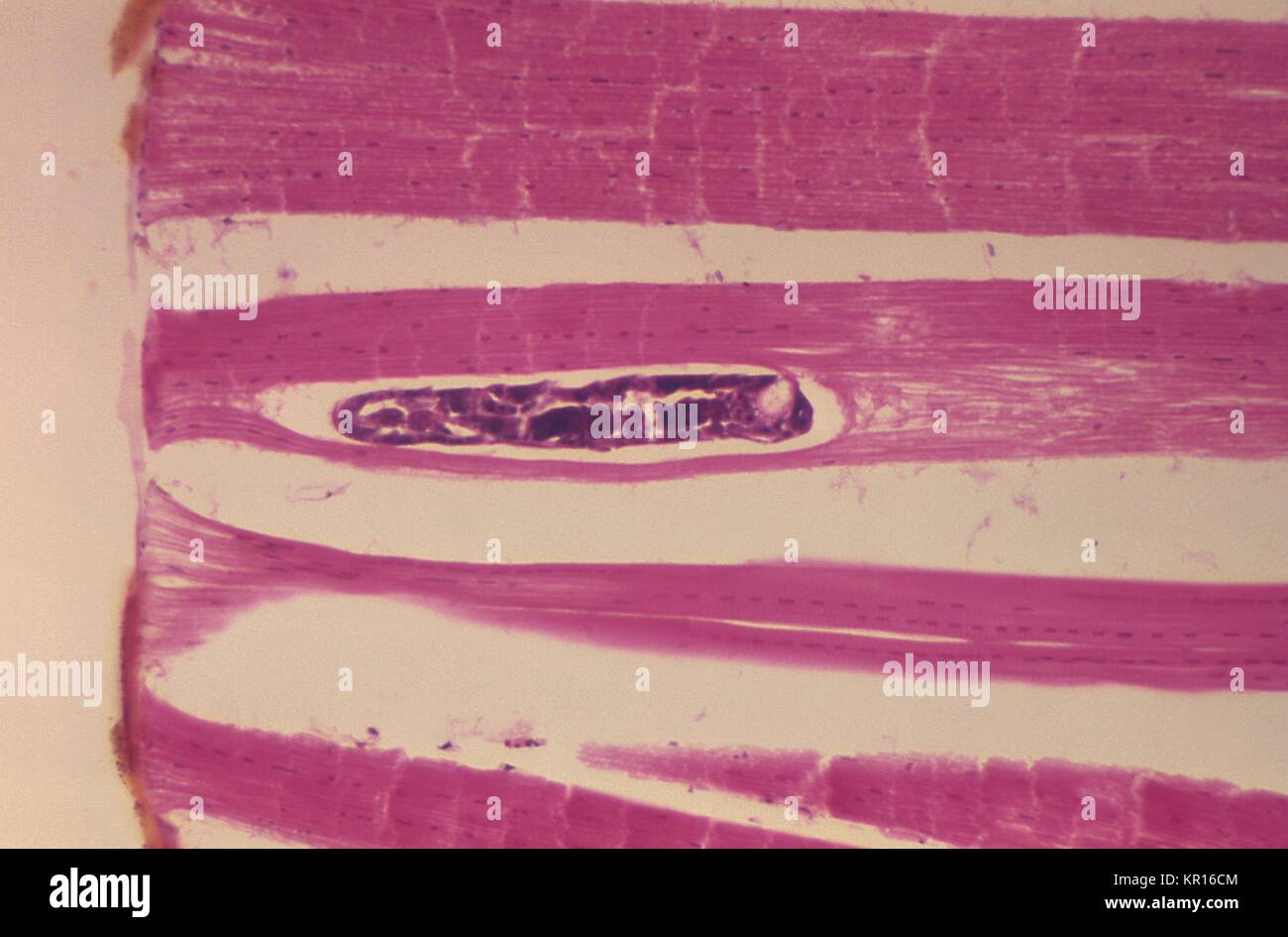
› 37006818 › Junqueiras_BasicJunqueira's Basic Histology Text and Atlas, 14th Edition Junqueiras Basic Histology Text and Atlas 14th Edition Vet Books ir. Zelle Peredas. Download Free PDF View PDF. Basic Histology. MARIAN ESTRADAR. Download Free PDF ... Solved Label the tissue and structures on this histology - Chegg Expert Answer From left to right … View the full answer Transcribed image text: Label the tissue and structures on this histology slide. 4 Intercalated disc Skeletal muscle fiber Skeletal muscle Cardiac muscle fibe Z disc Nucleus › cardiomyocytesCardiomyocytes (Cardiac Muscle Cells)- Structure, Function ... Microscopy (Histology) In order to observe cardiomyocytes under the microscope, it is necessary to fix and attach the cells on a microscope. Once the cells have been fixed and permeabilized on the slide, then they are ready for staining and viewing. Requirements A sample of cardiomyocytes (obtained from such an animal as rodents) Paraformaldehyde › articles › s41467/022/31339-8Instant diagnosis of gastroscopic biopsy via deep-learned ... Jul 13, 2022 · These advantages have enabled label-free histopathology of fresh tissue specimens, revealing key diagnostic histological features of various types of human solid tumors and diseases 14,15,16,17,18 ...
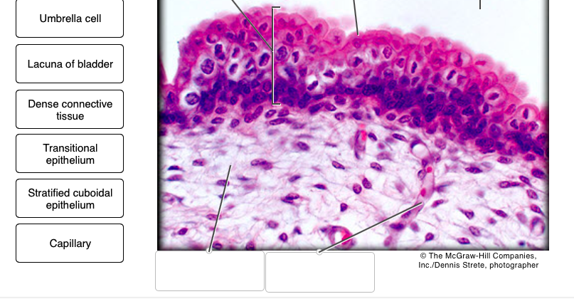
Histology: Labeling parts of tissues - Quizlet Histology: Labeling parts of tissues STUDY PLAY Nucleus, lumen, cell membrane What are the 3 things that have to be labeled in simple squamous epithelium? Lumen, basement membrane, nucleus, cell membrane What are the 4 things that need to be labeled in stratified squamous epithelium (keratinized=denser)? Lumen, nucleus, cell membrane › science › articleDeep neural network models for computational histopathology ... Jan 01, 2021 · Accurate segmentation of structures from histology images often requires the pixel-level delineation of object contour or the whole interior of the object of interest. CNNs trained to classify each patch centred on a pixel of interest as either foreground or background, can be used for segmentation tasks by employing a sliding-window approach. LAB- Connective Tissue (proper).pdf - HISTOLOGY of... The following headers will precede each histology slide: Lab: Loose Connective Tissues: below (Note: fibers are loosely packed in the matrix) Name of Tissue histologyguide.com Slidebox Text Figure areolar/loose connective tissue Label: collagen fiber elastic fiber fibroblast Ch 3 Connective tissue (Loose CT): MH 260 Ch 11, Skin MHS 277-279 ... Solved Label the tissues and structures on the histology - Chegg Label the tissues and structures on the histology slide. Squamous cell Nucleus of squamous cell Keratinized stratified squamous epithelium Simple squamous epithelium Stratified cuboidal epithelium Nonkeratinized stratified squamous epithelium Cuboidal cell The McGraw-Hill Companies, Inc./Al Telser, photographer Lamina propria Reset Zoom
Histology slide guide
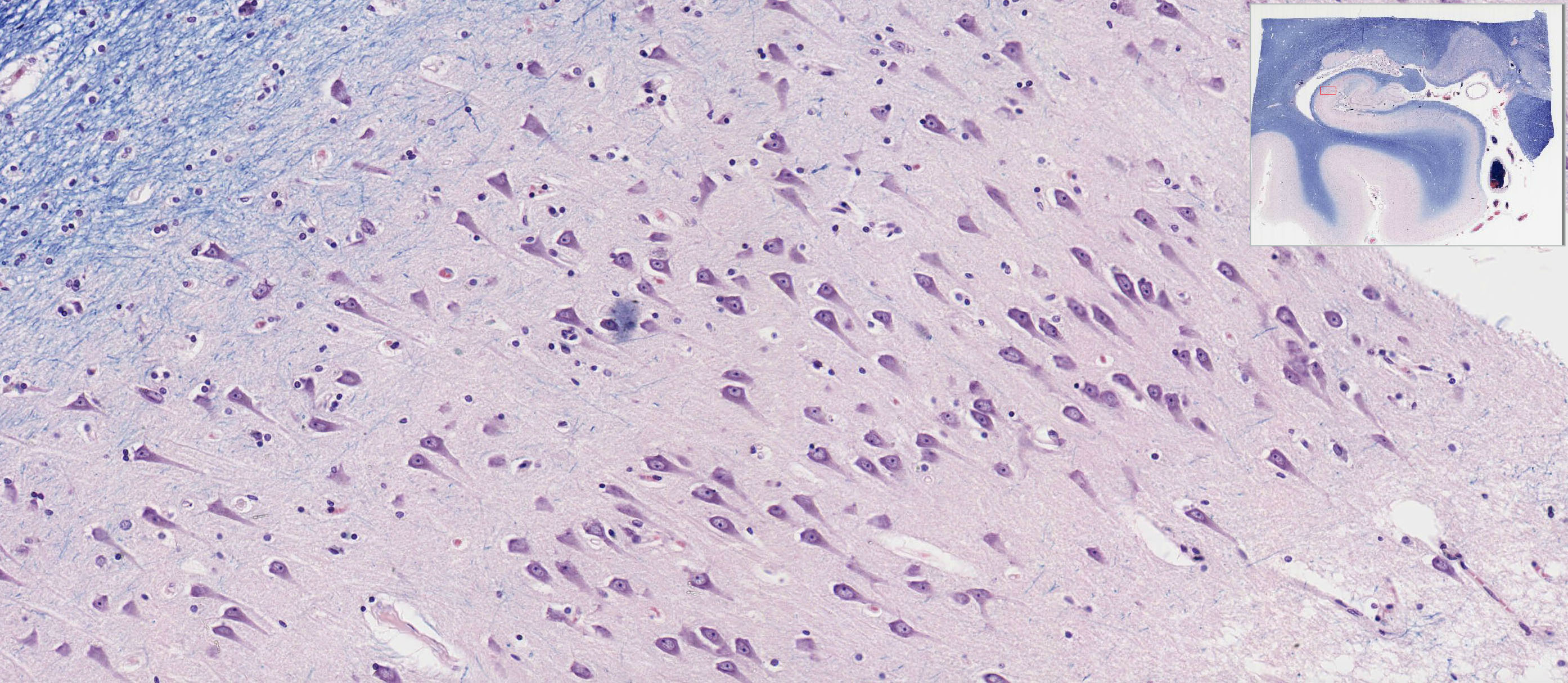
Cerebrum Histology - 6 Different Layers with Labeled Diagram So, this is a slide of cerebral cortex of animal brain. Layers of cerebrum histology with slide images and labeled diagram. Fine, do you want to know details about the different layers of cerebrum histology with real slide images? Okay, I am going to discuss on the two main parts of cerebrum structure separately. Histological features of ...
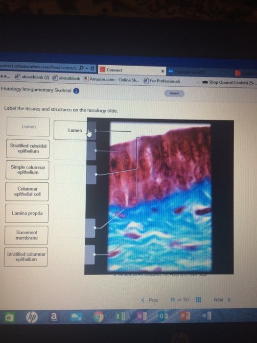
Solved] Label the tissues and structures on the histology ...
Labeling in Histology - Labtag Blog Labeling in Histology. Histology, the study of the anatomy of cells and tissues, is an important field of research used by researchers and physicians. While researchers seek to understand how each individual cell affects the function of tissues and organs, physicians study the histopathology of tissues, to see how they change in those affected ...

Solved Help Sa Chapter 5 Homework Assignment Label the ...
Introduction to histology - SlideShare • (C) batch will be having histology lab on Wednesday from 2.00-4.00pm. • The students have to bring the histology record, pink, blue and HB2 lead pencils. • They have to attend the prelab and study the slides under microscope and draw the tissue and label it. • Discuss the slide and identification points.

Caroline Thaung on Twitter: "Here's a cute little structure ...
How to examine histology slides: Techniques and tips | Kenhub How to examine histology slides Author: Alexandra Sieroslawska MD • Reviewer: Dimitrios Mytilinaios MD, PhD Last reviewed: November 26, 2021 Reading time: 3 minutes Histology is a beautiful subject that allows us to explore the structure and function of tissues. If we take a little sample of an organ tissue, stain it accordingly and place it under a light microscope, we are able to see the ...
Frontiers | Deep Learning for Whole Slide Image Analysis: An ...
tissue histology slides Flashcards | Quizlet Start studying tissue histology slides. Learn vocabulary, terms, and more with flashcards, games, and other study tools. ... dense regular connective tissue. areolar connective tissue. OTHER SETS BY THIS CREATOR ... inside and outside of heart 43 Terms. WhitneyGosvener. Structure of Heart 31 Terms. WhitneyGosvener. heart flashcards 46 Terms ...

31 Histology ideas | anatomy and physiology, histology slides ...
(Get Answer) - Label the structures on the tissue slide.. Label the ... A 406 x 178 UB 54 is simply supported at the ends of a span of5m and carries an uniformly distributed load of 60KN/m.Calculate the maximum deflection (E= 210000N/mm²) An inverted structural steel tee beam is sup ported by a pin at A and rests on a roller at B, as shown in Fig. P6.4-15a.

Pin by squashpotto on Anatomy | Anatomy, Electronic products ...
Slides of Histology | Anatomy and Physiology I | | Course Hero When we study smooth muscle and peripheral nerve tissue we will come back to this slide to try and distinguish between collagen fibers and fascicles of smooth muscle and/or nerve fibers and ganglia. NOTE: Slide 250 illustrates a point about the limits of classification schemes.
Solved] Label the tissues and structures on the histology ...
Histology Slides Flashcards | Quizlet Start studying Histology Slides. Learn vocabulary, terms, and more with flashcards, games, and other study tools. ... Structures: nucleus (none to label) Things to pay attention to: may see round structures, or long ones that are tubules cut on an angle ... protect underlying tissue Structures: flattened surface cells, basement membrane ...
Connective Tissue | Anatomy and Physiology | | Course Hero
› 36111379 › DiFiores_Atlas_ofDiFiore's Atlas of Histology with Functional Correlations ... veterinary histology module Sultan A Neja There is shortage of references in higher teaching institutions especially in newly opened institutions engaged in training of various Veterinary professionals in the country.

Histology of Blood Vessels
Histology Slides Identification from Different Organ Systems This article will show you histology slides from the following different organs system of an animal's body with identifying features. #1. Histology slide of epithelial tissue #2. General connective tissue histology slide #3. Histology slides of special connective tissue (blood, bone, and cartilage) #4. Muscular tissue histology slide #5.
Central Nervous System | histology
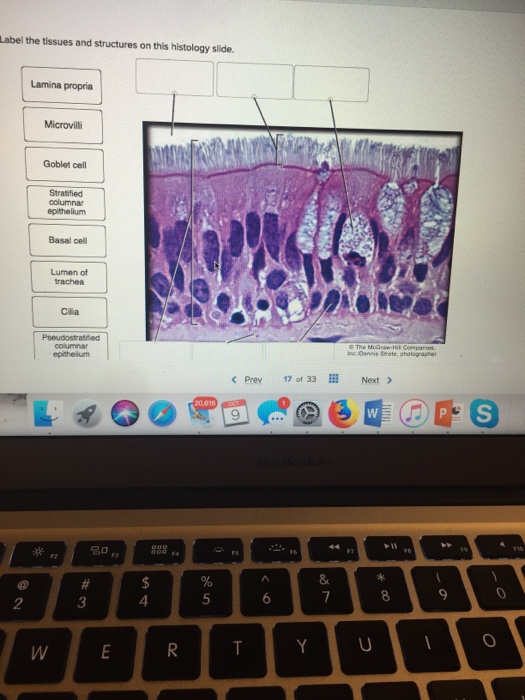
Solved Label the tissues and structures on this histology | Chegg.com Question: Label the tissues and structures on this histology slide. Lamina propria Goblet cell Stratified columnar epithelium Basal cell Lumen of Cilia The Mcüraw < Prev 170' 33Ⅲ Next > 名0 2 3 4 This problem has been solved! See the answer Label tissues and structures on this histology slide. Show transcribed image text Expert Answer
Microscope Slides of Cells and Tissues | Histology Guide
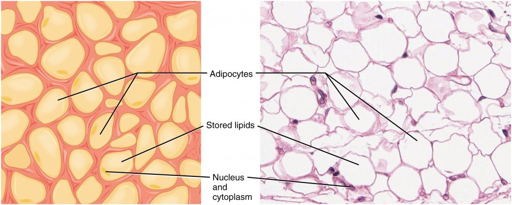
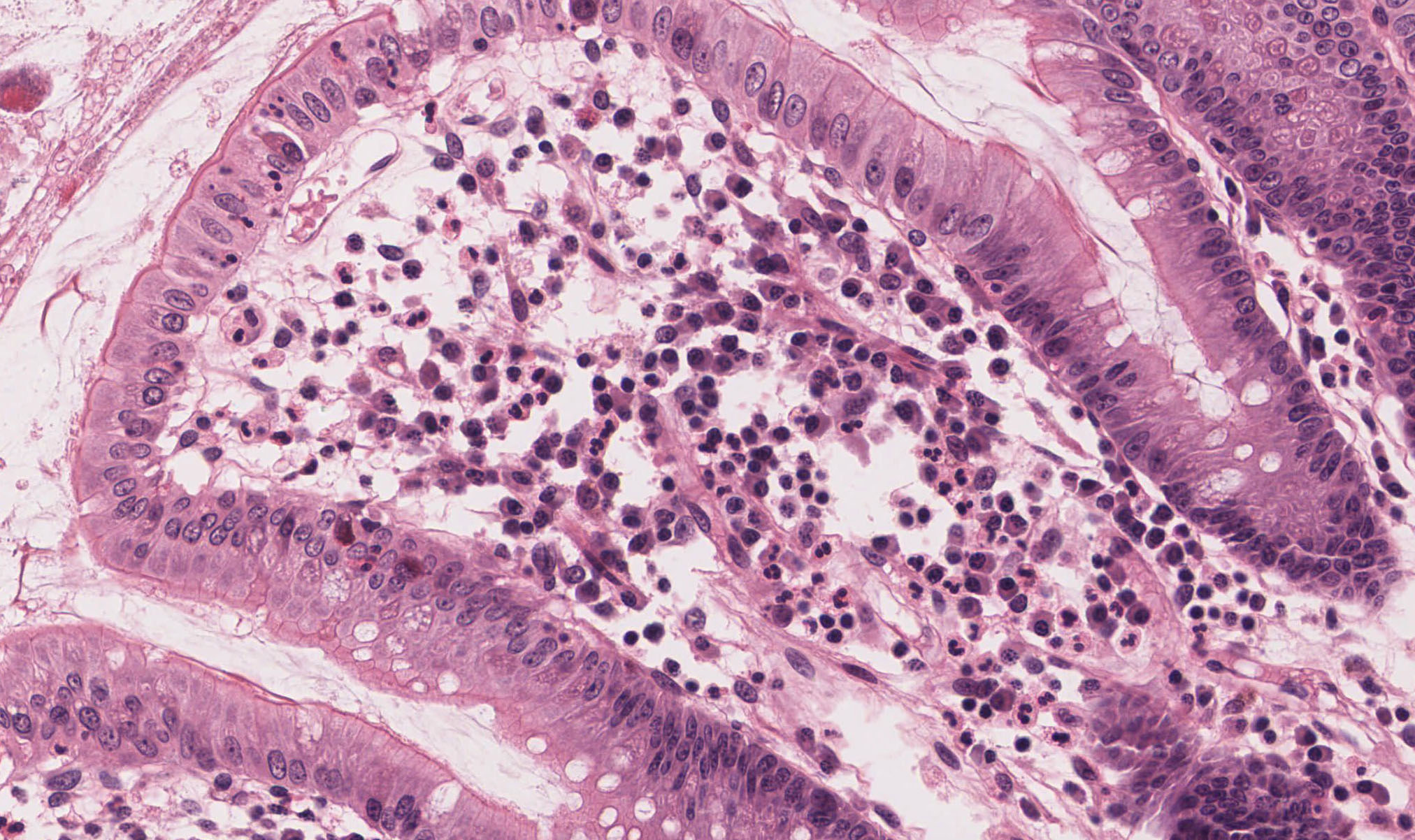
[Solved] Label the tissues and structures on the histology slide ... In the first slide, A. It is reticular connective tissue, as they are seen as a mesh work with high cellular structures. B. It is an adipose tissue, as the nucleus is seen at the periphery of the cell, with a thin rim of the cytoplasm C. Nucleus is seen in the periphery of the white adipose tissue. D. Lipids, are stored within large fat cells.
Atlas of Human Histology
label diagram of tissue cells label diagram of tissue cells Basic Histology -- Smooth Muscle, Longitudinal Section we have 9 Pics about Basic Histology -- Smooth Muscle, Longitudinal Section like Pseudostratified Ciliated Columnar Epithelium Shows Cilia Ciliated, Human Anatomy Lab Exercises Tissues Recognition and Function Flashcards and also Nervous Tissue - YouTube.

Using Histology Slides to Enhance Mammalian Dissection ...
histology human anatomy tissue slides - Quizlet epithelial tissue histology slides stratified squamous epithelium simple ciliated columnar epithelium pseudostratified ciliated columnar epit… flattened tile-like cells in surface layer, rounder cells in b… elongated cells, oval shaped nuclei, single layer, projections… Actually a single layer of cells of varying height, some not r… 11 sets Kenhub
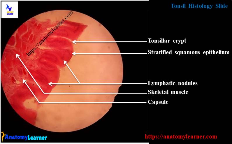
Tonsil Histology Slide with Labeled Diagram - Histological ...
Label the tissues and structures on the histology slide. - Transtutors Label the structures and tissues on this histology slide li (brush membrane Goblet cell Classify each of the following as acidic, basic, or neutral. Acidic Neutral Basic pH of 3 2:1 ratio of OH to H Pure water pH of 7 H concentration of 10 Soap...
Smooth muscle: Structure, function, location | Kenhub
Tissue Preparation Histology - SlideShare Importance Microscopic analysis of cells and tissues requires the preparation of very thin, high quality sections (slices) mounted on glass slides and appropriately stained to demonstrate normal and abnormal structures. • It involves several steps that are following. 1. Obtaining a fresh specimen 4. Clearing 2. Fixation 5. Wax infiltration 3.
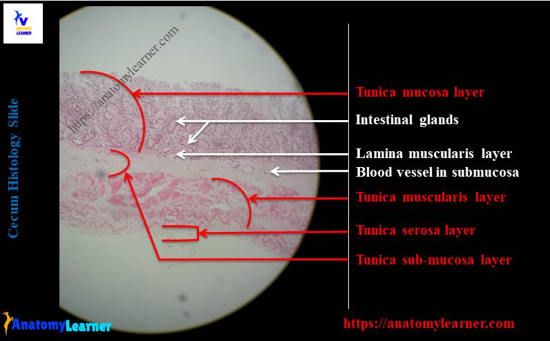
Cecum Histology Slide with Labeled Image and Diagram ...
Foundations - Histology Cells and Tissues - Embryology - UNSW Sites A tissue is a functional aggregation of similar cells and their intercellular materials that combine to perform common functions. An organ is an anatomically discrete structure (e.g. heart, skin) with 1 or more functions. Four tissues are considered basic or primary: epithelial, connective, muscular and nervous.

Histology Slides Flashcards | Quizlet
Histology guide: Definition and slides | Kenhub At a histological level, both the heart and blood vessels consist of three layers: Endothelial layer - epithelial tissue formed by simple squamous (endothelial) cells. In the heart, this layer is referred to as endocardium. Muscular layer - smooth muscle in the blood vessels, cardiac muscle (myocardium) in the heart.

Solved Label the tissue and structures on this histology ...
Microscope Slides of Cells and Tissues | Histology Guide This virtual slide box contains 275 microscope slides for the learning histology. Fig 023 Types of Tissue Cells and Tissues Tissues are classified into four basic types: epithelium, connective tissue (includes cartilage, bone and blood), muscle, and nervous tissue. Chapter 1 The Cell Chapter 2 Epithelium Chapter 3 Connective Tissue Chapter 4 Muscle

Lab Notes Home Page | Tissue biology, Histology slides, Loose ...
Skeletal Muscle Histology Slide Identification and Labeled Diagram ... From the skeletal muscle histology slide, you might identify the following important structures under the light microscope. Please try to find out these structures from the skeletal muscle slide labeled images. #1. Longitudinal section of skeletal muscle #2. Cross-section of skeletal muscle #3. Skeletal muscle fibers of the longitudinal section #3.

Endocrine Systems Lab
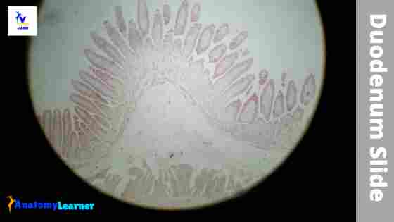
Duodenum Histology Slide with Labeled Diagram ...
Solved Label the the tissues and structures on the | Chegg.com
Solved Label the tissues and structures on this histology ...
Connective Tissue | histology

Endocrine Systems Lab

Histology of Blood Vessels
Solved Label the tissues and structures on the histology ...

Cartilage & Bone Quiz - Slide#1
Histology Slides, through Midterm

Microscopic view of the liver of feral pigeon of control ...

Histology Simple Columnar Epithelial Tissue | Histology ...

Lab.02 Bio.141: Histology (virtual slides) Flashcards | Quizlet

🔴 Liver abscess and cirrhosis. hepatic duct. Bile duct of liver pathology.
A&P I Lab | Exercise 4: Histology & Tissues

Teaching Resource Tongue Human Histology Tissue Prepared Slides For Microscopes - Buy Teaching Resource,Human Histology Slides,Prepared Slides Product on Alibaba.com
Pharynx, Esophagus, and Stomach | histology
Virtual histological staining of label-free total absorption ...

Tendon histology slides (H&E 40X).: (A) Longitudinal section ...
A photomicrograph revealing the histology of Onchocerca ...

adipose tissue | Vet medicine, Adipose tissue, Histology slides

Tissues Chapter 5. Name the tissue Name a structure it is ...
HA 235 - Histology - Female Reproductive System
Histology Tutor
Post a Comment for "44 label the tissue and structures on this histology slide"