39 dental x-ray tube head diagram
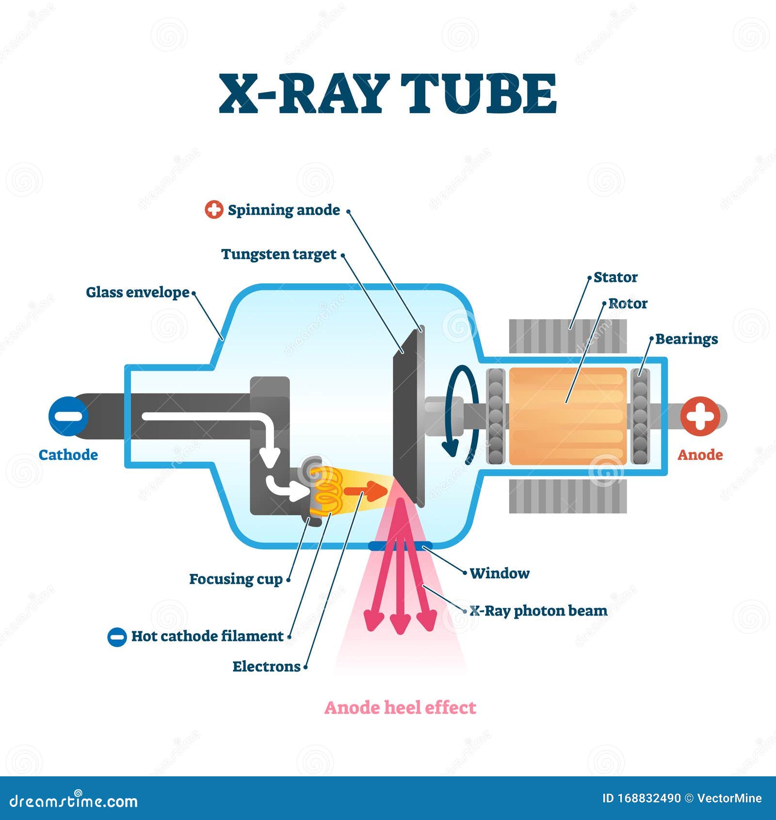
Home | Hamamatsu Photonics The official website of Hamamatsu Corporation whose mission is to advance science and industry through photonic technologies. Our products include optical sensors and components, cameras, light & radiation sources, lasers, and customized solutions. X-ray Machine Parts and Functions Explained - Uni X-ray An x-ray tube primarily functions as an energy converter that receives energy and converts it into two other different forms of energy, x-ray radiation energy, and heat energy. II. X-ray Detector. X-ray detectors refer to instruments deployed in measuring the spatial distribution, flux, spectrum, and every other x-ray property.
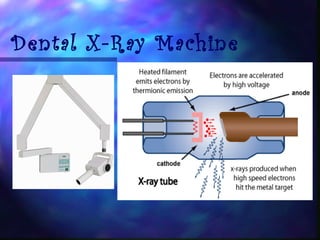
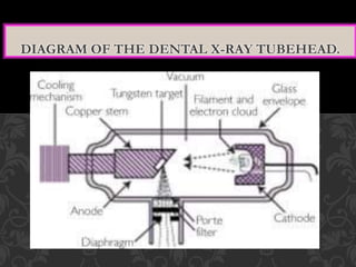
LECTURE 1: The Tube Head - Intro Dental Radiography I LECTURE 1: The Tube Head The Dental X-ray Tube The filament is heated by the filament current. Electrons are emitted by the hot filament and travel to the anode. This flow of electrons from the...
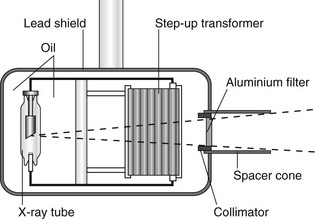
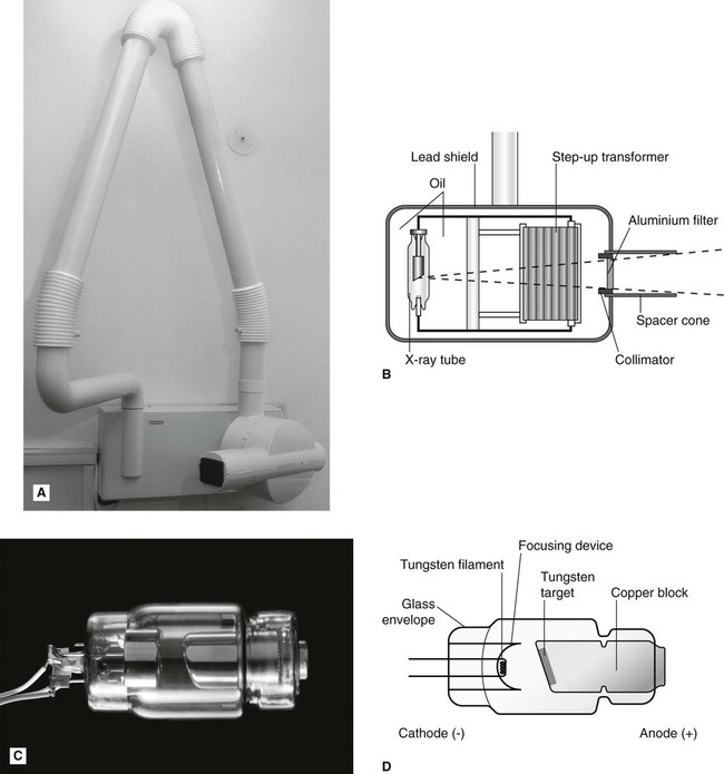
Dental x-ray tube head diagram
Safety Code 35: Safety Procedures for the Installation, Use ... However, in systems where an X-ray tube for radiography is also present, the shielding for this X-ray tube must be evaluated independently, as in Section B1.3.2. When equipment include more than one X-ray tube, such as in cardiac systems, the shielding calculation must take into account each X-ray tube independently. Simplified Positioning for Dental Radiology - Dentalaire Products Dental X-Ray, X-Ray Products. DTX Digital Dental Imaging System; DTX Dental CR Reader; ... Tip the tube head 45 degrees to the side of the face. ... These diagrams are best used in conjunction with the positioning "Cheat Sheet" above. When performed as indicated, these simple positioning guidelines will provide well-positioned dental films. Types of Dental X-Rays and Why You Need Them A dental x-ray is the common term for a dental radiograph. It is one of the dentist's most important diagnostic tools, giving him or her a better picture of what's going on with your teeth than simply looking in your mouth. Dental radiographs work by using a small, controlled burst of radiation to create a picture of the tooth.
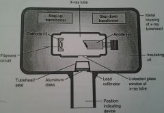
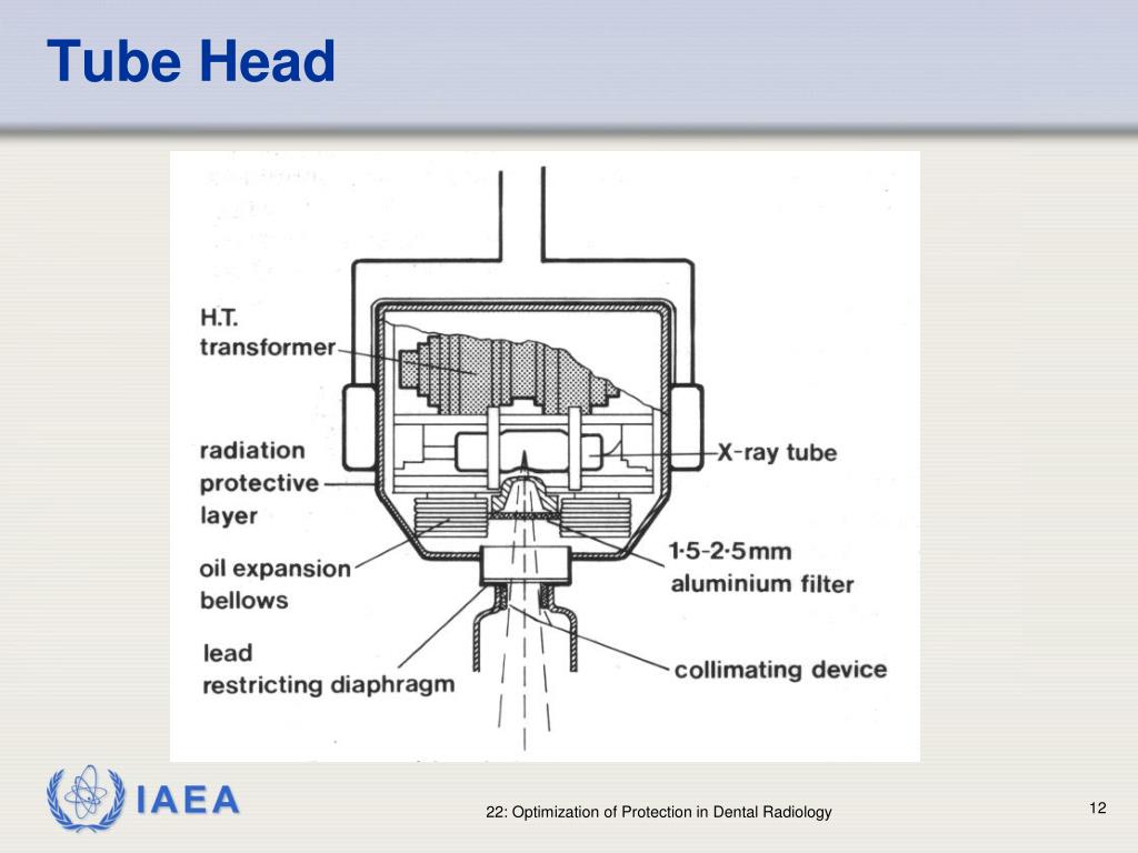
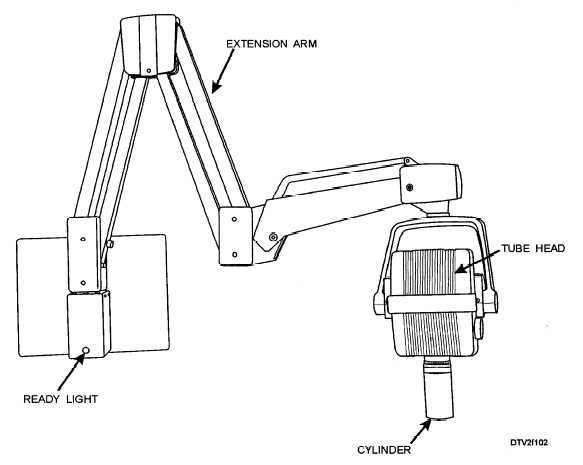
Dental x-ray tube head diagram. Labelling Dental X-Ray Tube head Diagram | Quizlet Start studying Labelling Dental X-Ray Tube head. Learn vocabulary, terms, and more with flashcards, games, and other study tools. Scheduled maintenance: Saturday, June 5 from 4PM to 5PM PDT How does a dental x ray tube head work? - Answers Inside the metal tube housing is the x-ray tube. The diagram in figure 1-2 represents a dental x-ray tube head and a dental x-ray tube. This tube emits radiation in the form of photons (photons... Dental X-ray Tube Head Diagram - local dentist Dental X-ray Tube Head Diagram. The dental x-ray technician should never receive primary radiation from a dental The following diagram will identify the location of these two devices (see figure 1-8). Tube head assembly: filter, collimator (diaphragm), PID or cone or tube. PDF Easy Guide to Dental X-ray Positioning - AAHA Ai m at the area between finger and thumb. Line up the bottom line on the tubehead to the canine tooth 103-101 & 203-201 (R or L incisors) 45 -50° Dependent upon the headtype of patient - tube head parallel to the nose crack 103-203 (All incisors) 45-60° Parallel to nose crack Mandible
The Functions of an X-Ray Tube - The X-Ray Tube It absorbs isotropic (direction-independent) x-ray photons hence reducing the leakage of radiation. It is also acts to concentrate the electrons so they escape through the little hole that has been created in the bottom of the shield. Two blue straws. Anode. Radiation Protection - Welcome to Dental Radiography The following diagram will identify the location of these two devices (see figure 1-8). Figure 1-8. Tube head assembly: filter, collimator (diaphragm), PID or cone or tube. b. Filter. The aluminum filter or disk is placed in the path of the x-ray beam. Figure 1-8 shows the location of the PID. Production of Dental X-Rays - Dental Radiology - YouTube A summary of how an xray is produced in the dental xray tube head.Information taken from:Dental Radiography Principle and Techniques - by Iannucci and Howert... Dental X-ray Tube Head How It Works - local dentist Dental X-ray Tube Head How It Works. The tubehead is a sealed, heavy metal housing that contains the x-ray tube that has a specific function that contributes to the safe exposure of dental x-rays. the safety features, will allow you to explain to the patient how x-rays work. Dental X-ray Tube Head How It Works. How does the procedure work?
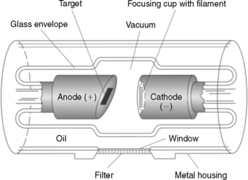
The X-ray Tube | Radiology Key The general-purpose x-ray tube is an electronic vacuum tube that consists of an anode, a cathode, and an induction motor all encased in a glass or metal enclosure (envelope). Figure 5-3 provides a labeled illustration of this design. Recall that the anode is the positive end of the tube and the cathode is the negative end of the tube. US4157476A - Dental X-ray tube head - Google Patents In a dental x-ray tube head, the x-ray tube is in a casing that supports the tube and shields against stray radiation being projected to the environment through the housing of the tube head. The... Cathode (x-ray tube) | Radiology Reference Article - Radiopaedia The cathode is part of an x-ray tube and serves to expel the electrons from the circuit and focus them in a beam on the focal spot of the anode.It is a controlled source of electrons for the generation of x-ray beams. The electrons are produced by heating the filament (Joule heating effect) i.e. a coil of wire made from tungsten, placed within a cup-shaped structure, a highly polished nickel ... 3: Dental X-ray equipment, image receptors and image processing Fig. 3.2 Diagram of the tubehead of a typical dental X-ray set showing the main components. • The glass X-ray tube, including the filament, copper block and the target (see Ch. 2) • The step-up transformer required to step-up the mains voltage of 240 volts to the high voltage (kV) required across the X-ray tube
PDF INSTALLATION INSTRUCTIONS - Belmont Equipment x-ray tube, high voltage generator or both. B. Duty cycle A cool down interval of 50 seconds or more must be allowed between each 1 second exposure. (a 25 second cool down must be allowed between each 0.5 second exposure.) This will avoid the accumulation of excess heat and prolong the tube head life. C. Tube head cooling curve 0 0
How to Build an X-ray Machine | Science Project Option 1: Mount the power supply and X-ray tube on a wooden board as shown in Figure 3. This option allows the X-ray machine to be easily transported and is the option we will continue to depict in subsequent images. Mount the power supply using Velcro; secure the X-ray tube by feeding the attached wires though slots in the wooden mount.
Steps In The Process Of Xray Production - Dental Radiography See figures 1-3, 1-4, and 1-5 for a diagram of the complete procedure. Figure 1-3. Tube head with the filament of the cathode emitting electrons. Figure 1-4. Electrons speeding toward the anode (tungsten target). Leaded Glass Tube Metal Housing Cathode Anode (Tungsten Target) Cathode Anode (Tungsten Target) Aluminum Filter
PDF Intraoral radiographic techniques The Periapical radiograph (IOPA) is the basic investigation that gives graphic information about the alveolar bone, periodontal areas and the hard tissues of the tooth. Each image usually shows 2-4 teeth. Indications The clinical indications include: 1. To visualize Periapical region 2. detection of apical infection/inflammation. 3.
Facial trauma - Wikipedia Tracheal intubation (inserting a tube into the airway to assist breathing) may be difficult or impossible due to swelling. Nasal intubation, inserting an endotracheal tube through the nose, may be contraindicated in the presence of facial trauma because if there is an undiscovered fracture at the base of the skull, the tube could be forced ...
Panoramic Dental X-ray - Radiologyinfo.org Panoramic radiography, also called panoramic x-ray, is a two-dimensional (2-D) dental x-ray examination that captures the entire mouth in a single image, including the teeth, upper and lower jaws, surrounding structures and tissues. The jaw is a curved structure similar to that of a horseshoe.
Production Of X-Rays - Welcome to Dental Radiography Inside the metal tube housing is the x-ray tube. The diagram in figure 1-2 represents a dental x-ray tube head and a dental x-ray tube. This tube emits radiation in the form of photons (photons will be discussed in Lesson 2) or x-rays. X-ray photons expose the film. In addition to exposing the film, it also exposes the patient to radiation.
Dental X-ray tube head - General Electric Company Dental x-ray apparatus which includes the new shielding construction is depicted in FIG. 1. The dental x-ray tube head is generally designated by the reference numeral 10. It comprises a housing 11 having a bottom wall 12 to which a tubular assembly 13 is attached. This assembly is otherwise known as a cone.
Can a Sinus Infection Be Caused by a Tooth? | Oral Answers Here is an x-ray of a tooth that had a root canal and crown done previously, but the infection at the roots had never quite healed. I have outlined some of the important structures below for those of you who are not accustomed to reading x-rays. I colored the sinus blue and the tooth infection red in the x-ray below:
Dental X-ray Tubehead Diagram | Quizlet piece of lead that reshapes the size of the beam and further filters out low-wavelength beams PID Position Indicator Device: device that is lined with lead and contains an aperture through which the primary x-ray beam passes as it leaves the device and heads to the patient Copper Stem (anode) positive electrode Focusing Cup (cathode)
PDF DENTAL X-RAY 097 - Belmontdental the x-ray tube, high voltage generator or both. B. Duty cycle A cool down interval of 50 seconds or more must be allowed between each 1 second exposure. (a 25 second cool down must be allowed between each 0.5 second exposure.) This will avoid the accumulation of excess heat and prolong the tube head life. C. Tube head cooling curve 1.
Label the Dental X-ray Tubehead (Screencast) - Wisc-Online OER Label the Dental X-ray Tubehead (Screencast) By Joan Rohrer The tubehead is a sealed, heavy metal housing that contains the x-ray tube that produces dental x-rays. This learning object will provide students with practice identifying and labeling the dental x-ray tubehead. Urine Colony Counts By Kristine Snow Learners watch a brief video clip.
Intraoral X-ray unit | Planmeca ProX Planmeca ProX™Flexible intraoral X-ray unit. Planmeca ProX™. The advanced Planmeca ProX™ intraoral X-ray unit provides easy and precise positioning, a straightforward imaging process and top-quality images in high resolution. It is a highly beneficial and effective 2D imaging option for all dental clinics.
X-ray tube - Wikipedia An X-ray tube is a vacuum tube that converts electrical input power into X-rays. The availability of this controllable source of X-rays created the field of radiography, the imaging of partly opaque objects with penetrating radiation.In contrast to other sources of ionizing radiation, X-rays are only produced as long as the X-ray tube is energized.X-ray tubes are also used in CT scanners ...
Top Dental Digital Radiography Mistakes - DentalSensors.com Then make sure your x-ray head tube is flush against the ring. There should be less than an inch gap between the end of the x-ray head tube and the patients skin. X-ray head generators are a lot like a shot gun. The farther you are away from your target or in your case a dental sensor. The less you are going to hit that target.
Glass tubing function - tkymmq.farmbridge.shop Aug 02, 2017 · OLYCRAFT 16 Pack Laboratory Glass Tube Borosilicate Glass Tube Clear Glass Borosilicate Blowing Tubes 12mm OD 1.5mm Thick Wall Tubing 4 Inch Long Clear Tube with Nylon Tube Pipe Brushes. 4.4 out of 5 stars 22. $13.29 $ 13. 29 ($0.83/Item) Get it as soon as Sat, Jul 23.
Types of Dental X-Rays and Why You Need Them A dental x-ray is the common term for a dental radiograph. It is one of the dentist's most important diagnostic tools, giving him or her a better picture of what's going on with your teeth than simply looking in your mouth. Dental radiographs work by using a small, controlled burst of radiation to create a picture of the tooth.
Simplified Positioning for Dental Radiology - Dentalaire Products Dental X-Ray, X-Ray Products. DTX Digital Dental Imaging System; DTX Dental CR Reader; ... Tip the tube head 45 degrees to the side of the face. ... These diagrams are best used in conjunction with the positioning "Cheat Sheet" above. When performed as indicated, these simple positioning guidelines will provide well-positioned dental films.
Safety Code 35: Safety Procedures for the Installation, Use ... However, in systems where an X-ray tube for radiography is also present, the shielding for this X-ray tube must be evaluated independently, as in Section B1.3.2. When equipment include more than one X-ray tube, such as in cardiac systems, the shielding calculation must take into account each X-ray tube independently.




![PDF] Tube angulation effect on radiographic analysis of the ...](https://d3i71xaburhd42.cloudfront.net/2474d7c2e22cba363573626935ca08ecc19e7744/3-Figure2-1.png)






Post a Comment for "39 dental x-ray tube head diagram"