44 circle of willis kenhub
Circle of Willis: Anatomy and function | Kenhub The circle of Willis (cerebral arterial circle or circulus arteriosus) is an anastomotic ring of arteries located at the base of the brain. This arterial anastomotic circle connects the two major arterial systems to the brain, the internal carotid arteries and the vertebrobasilar (vertebral and basilar arteries) systems. It is formed by four paired vessels and a single unpaired vessel with numerous branches that supply the brain. Vascular Workbook p 5-8, Extracranial & Intracranial Anatomy Define the term "fetal origin of the PCA" used to describe one type of anatomic variation of the Circle of Willis. ... Spinal Cord - Neuroanatomy | Kenhub Anatomy Guide. Kenhub. $13.99. STUDY GUIDE. Vascular 2, Intracranial & Extracranial Anatomy 92 Terms. studentmomof3. Anatomy of the thorax Q&A 264 Terms. sabir_khan2.
Circle of Willis quizzes and unlabeled diagrams | Kenhub Simply(ish) put, the circle of Willis is a circulatory anastomosis supplying blood to the brain and neighboring structures. These arteries are located at the base of the brain, close to the optic chiasm. Circle of Willis arteries. The circle of Willis, also known as the cerebral arterial circle, is formed by anterior and posterior arterial pathways. The arteries of the circle of Willis include:

Circle of willis kenhub
› en › libraryAnterior cerebral artery: Anatomy, branches, supply | Kenhub Aug 04, 2022 · This anastomosis makes the anterior/rostral component of the circle of Willis, which is the most important anastomosis between the cerebral vessels. The other arteries that comprise the Willis' circle are the internal carotid, posterior cerebral, anterior communicating and posterior communicating arteries. Willis Polygon: Location, Anatomy, and Functions | Life Persona He Willis's polygon , Also called the Willis ring or cerebral arterial circle, is an arterial structure in the form of a heptagon that is located at the base of the brain. This structure is formed by two groups of arteries: the internal carotid arteries and the vertebrobasilar system. anatomy cerebral des veins Bloodsupplyofthebrain 101102134629-phpapp02. 17 Pictures about Bloodsupplyofthebrain 101102134629-phpapp02 : Veins of the Brain - Anatomy and Clinical Notes | Kenhub, Cerebral Veins - Earth's Lab and also Chapter 23 Cardiovascular system - Biology 4 Human AnatomyProfessor. Bloodsupplyofthebrain 101102134629-phpapp02
Circle of willis kenhub. Meninges, Ventricles, CSF and brain blood supply | Kenhub The circle of Willis, officially termed the 'cerebral arterial circle', is a hexagonal anastomotic vascular network at the base of the brain. It has two main sources. It has two main sources. The first are the two internal carotid arteries and their branches - anterior and middle cerebral arteries. Circle of Willis - 3D Anatomy Tutorial - YouTube 3D anatomy tutorial on the Circle of Willis.-----Join me on Instagram: : ... Full text of "Télé Poche - No. 2776 - 22-04-2019" An icon used to represent a menu that can be toggled by interacting with this icon. anatomy of veins and arteries - Microsoft Anatomy arteries and veins. Anatomy groin neurovascular vascular femoral arteries illustration pelvis artery veins vein nerve medivisuals1 system circulatory saphenous medical superficial. Veins arteries anatomy human body system circulatory artery guide arterial vessels study medicinebtg lab sm pulmonary wpclipart credit.
Circle of willis : Anatomy 17.1k members in the Anatomy community. Press J to jump to the feed. Press question mark to learn the rest of the keyboard shortcuts Blood supply to the brain: Anatomy of cerebral arteries | Kenhub Circle of Willis is indeed a hot neuroanatomy topic! Master it with our circle of Willis quizzes & unlabeled diagrams. The circle of Willis is a polygonal structure that surrounds the optic chiasm and infundibulum, as it rests within the chiasmatic and interpeduncular cisterns. The anastomosis provides an alternative route for blood flow in the event of vascular occlusion. Hypothalamus - Anatomy, Blood supply and Function | Kenhub The anterior and posterior anastomoses form a circular arterial system known as the cerebral arterial circle of Willis. Anterior and posterior branches of the circle of Willis provide arterial blood to the hypothalamus. The hypothalamus also receives arterial supply from the hypothalamic branches of the superior hypophyseal artery. major veins and arteries Circle of willis 【 note -: major contribution of branches of internal. ... lung hilum anatomy right pulmonary pulmonalis artery bronchial kenhub lymph nodes phd md arteria reviewer dimitrios reviewed alice last nodi. Arteries Of The Right Upper Limb And Thorax Quiz . arteries upper limb thorax right quiz.
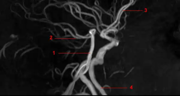
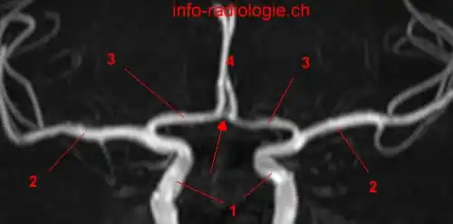
MRA of the Circle of Willis - W-Radiology The circle of Willis is where several arteries in the brain meet or join together (1). Also known as the circulus arteriosus cerebri or the cerebral arterial circle, the circle of Willis is an anastomotic (connecting) ring of arteries found at the base of the brain (2). Arteries of the brain: inferior view (preview) - Human Anatomy | Kenhub Arteries supply the brain with oxygenated blood. They include those that have close connections with one another to form networks, for example the circle of ... pic of anatomy of brain CTA of Circle of Willis - Neuro Case Studies - CTisus CT Scanning we have 8 Images about CTA of Circle of Willis - Neuro Case Studies - CTisus CT Scanning like brain, Anatomy, Medical, Head, Skull, Digital, 3 d, X ray, Xray, Image | Radiopaedia.org and also Facial vein: Anatomy, tributaries, drainage | Kenhub. Here you go: CTA Of Circle Of ... Circle of Willis | Radiology Reference Article | Radiopaedia.org The Circle of Willis is an arterial polygon (heptagon) formed as the internal carotid and vertebral systems anastomose around the optic chiasm and infundibulum of the pituitary stalk in the suprasellar cistern.
› en › libraryHypothalamus: Anatomy, nuclei and function | Kenhub Jun 28, 2022 · The anterior and posterior anastomoses form a circular arterial system known as the cerebral arterial circle of Willis. Anterior and posterior branches of the circle of Willis provide arterial blood to the hypothalamus. The hypothalamus also receives arterial supply from the hypothalamic branches of the superior hypophyseal artery.
› posterior-communicatingPosterior Communicating Artery: Anatomy, Function The posterior communicating artery (PCOM) is a part of a group of arteries in the brain known as the circle of Willis. The artery connects the internal carotid and the posterior cerebral arteries. Its role is to provide blood supply to the brain. The posterior communicating artery is a location where aneurysms can potentially occur.
Posterior communicating artery: Anatomy, function | Kenhub The posterior communicating artery in the circle of Willis The posterior communicating artery originates from the intracranial portion of the internal carotid artery, specifically from its C7 segment. The left and right posterior communicating arteries usually differ in size, with one of them being notably larger than the other.
Cerebrovascular Anatomy: Extracranial Anatomy and Circle of Willis ... Start studying Cerebrovascular Anatomy: Extracranial Anatomy and Circle of Willis. Learn vocabulary, terms, and more with flashcards, games, and other study tools.
1. The Cranial Nerves and the Circle Of Willis This connection, plus the connection between the internal carotid and vertebrobasilar systems, forms the Circle of Willis. This circle of arterial anastomoses provides a margin of safety should one of the major arteries be obstructed. Veins of the brain ( fig 1d) drain into large collecting channels in the dural folds called dural sinuses.
anatomy of spinal nerves willis circle cerebral arterial neuroanatomy neurosurgicalatlas correlation surgical. Foot, Plantar Surface (superficial) - Human Body Help ... cranial motor nerve accessory nerves kenhub anatomy accessorius anterior ventral. Frog nervous system. Cerebral arterial circle (circle of willis). Foot, plantar surface (superficial) - human body ...
Arteries of the brain: Posterior circulation | Kenhub The completed structure is known as the circle of Willis . Circle of Willis (caudal view) It surrounds the optic chiasm and infundibulum, as it rests within the interpeduncular cistern. The circular anastomosis was initially believed to provide alternative route for blood flow in the event of vascular occlusion.
Cardiovascular System Lab-circle of willis Questions and Study Guide ... circle of willis Terms in this set (7) Vertebral arteries these major arteries of the neck branch from the subclavians and merge to form the basilar midline Basilar artery this artery arises from the vertebral arteries at the junction between the medulla oblongata and pons.
Brain Anatomy - Physiopedia At birth, the average brain weighs about 350 - 400grams, approximately 25% of the final adult brain weight of 1.4 - 1.45 kg and accounting for only 2% of overall body mass, which is reached between 10 and 15 years of age. Fastest growth occurs during the first 3 years of life, with almost 90% of the adult value reached by the age of 5 years.
Cerebral Arterial Circulation: 3D Augmented Reality Models and 3D ... The circle of Willis ( Figure 1) is a ring of vessels that provides important colligative communications between the anterior and posterior circulations of the midbrain and hindbrain.
Circle of Willis Flashcards | Quizlet Start studying Circle of Willis. Learn vocabulary, terms, and more with flashcards, games, and other study tools. Search. Create. Log in Sign up. Log in Sign up. Circle of Willis. ... Kenhub Anatomy Guide. Kenhub. $18.99. STUDY GUIDE. Master Blood Vessels 77 Terms. Xaeromancer. Lab24_Aaron 51 Terms. PatrickNguyenDDS PLUS. Respiratory anatomy ...
Internal carotid artery: Anatomy, segments and branches - Kenhub The internal carotid arteries are part of the anterior circulation, which is responsible for supplying the forebrain. The two circulations of the brain anastomose and form an anatomical structure called the circle of Willis. Why are there two circulations and so many sources of arterial blood to the brain?
spleen and pancreas anatomy . cervical platysma muscles fascias kenhub deep anatomy fascia neck superficial facial anterior muscle fascial layers layer function located investing major. CTA Circle Of Willis - Neuro Case Studies - CTisus CT Scanning . ct willis circle scan cta ctisus neuro diagnosis studies case
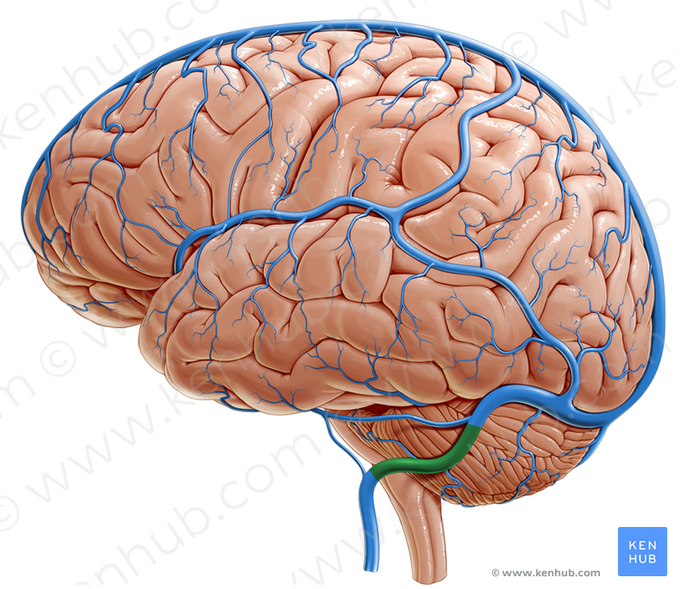
anatomy cerebral des veins Bloodsupplyofthebrain 101102134629-phpapp02. 17 Pictures about Bloodsupplyofthebrain 101102134629-phpapp02 : Veins of the Brain - Anatomy and Clinical Notes | Kenhub, Cerebral Veins - Earth's Lab and also Chapter 23 Cardiovascular system - Biology 4 Human AnatomyProfessor. Bloodsupplyofthebrain 101102134629-phpapp02
Willis Polygon: Location, Anatomy, and Functions | Life Persona He Willis's polygon , Also called the Willis ring or cerebral arterial circle, is an arterial structure in the form of a heptagon that is located at the base of the brain. This structure is formed by two groups of arteries: the internal carotid arteries and the vertebrobasilar system.
› en › libraryAnterior cerebral artery: Anatomy, branches, supply | Kenhub Aug 04, 2022 · This anastomosis makes the anterior/rostral component of the circle of Willis, which is the most important anastomosis between the cerebral vessels. The other arteries that comprise the Willis' circle are the internal carotid, posterior cerebral, anterior communicating and posterior communicating arteries.




:watermark(/images/watermark_only_sm.png,0,0,0):watermark(/images/logo_url_sm.png,-10,-10,0):format(jpeg)/images/anatomy_term/rami-terminales-superiores-et-inferiores-arteriae-cerebri-mediae/pFz2dcR50QWqqpChQZMw_Rr._terminales_superiores_et_inferiores_01.png)
:background_color(FFFFFF):format(jpeg)/images/library/3939/m97KU1MomTikfQ3g4Pn4QQ_A._carotis_interna_02.png)

:watermark(/images/watermark_only_sm.png,0,0,0):watermark(/images/logo_url_sm.png,-10,-10,0):format(jpeg)/images/anatomy_term/inferior-hypophysial-artery/GVgjzkB1CCsqr6RDpiCA_Inferior_hypophyseal_artery.png)

:background_color(FFFFFF):format(jpeg)/images/library/13938/qNRQ5TN7dGARxv8Qr5ysNg_Circulus_arteriosus_cerebri_01.png)
:background_color(FFFFFF):format(jpeg)/images/library/3940/uPLjNLzwRVOsHURrgBlGA_A._carotis_interna_01.png)


:background_color(FFFFFF):format(jpeg)/images/library/13734/middle-cerebral-artery_english.jpg)


:format(jpeg)/images/team_member/KH_image.png)

:background_color(FFFFFF):format(jpeg)/images/library/11763/labeled_diagram_circle_of_willis.jpg)




:watermark(/images/watermark_only_sm.png,0,0,0):watermark(/images/logo_url_sm.png,-10,-10,0):format(jpeg)/images/anatomy_term/internal-carotid-artery-6/J5VGRlbbchOeB7Rcedqog_internal-carotid-artery-6.png)



Post a Comment for "44 circle of willis kenhub"